Case study: The Labrador 'bringing up her food'

This case study follows a 3 year old dog who has been bringing up her food and feeling weak... can you solve it?

feature-img

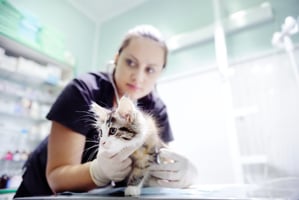
It's coming to the end of the day on a Thursday, and you're on clinics. Next on the list is a young Labrador who is vomiting....

Grab a pen and paper, set your timer (to record your CPD!) and settle down to solve the case... 

History and presenting problem

A 3-year-old female neutered Labrador Retriever presents to your practice because, in the past week, she has started ‘bringing up her food’. The owner also mentions that her bark has changed recently and that she is drooling more than usual. They also state that their dog seems a bit quieter and weaker than usual. On further questioning the owner reports that there is no abdominal effort when she is bringing her food up, and what she produces is largely undigested. This makes you suspicious that the dog is regurgitating rather than vomiting. 

Your clinical exam

Your physical examination reveals a normal mentation but a decreased palpebral reflex. Mucous membranes are pale pink with a capillary refill time of 1.5 seconds. The dog’s heart rate is 112 beats per minute, and they have a respiratory rate of 36 breaths per minute. Abdominal palpation is unremarkable and rectal temperature is 38.8C. The dog’s peripheral lymph nodes are within normal limits and there is no evidence of joint pain. You decide to trot the dog up and down outside the practice to assess her mobility. While she starts well and seems keen to walk, she soon becomes weak and struggles.

What is your problem list for this dog? 
Try to list each of the problems you've spotted in your history and clinical exam - this will help to ensure you don't miss any potential differential diagnoses later. When you are ready, click 'see more' to see the answers.

Regurgitation
Exercise induced weakness
Drooling
Change in vocalisation
Subdued demeanour
See more See less


What are your differential diagnoses for each of these problems?

Here are a few possible differentials (you may have come up with additional ones):

Regurgitation
– primary (idiopathic) megaoesophagus, secondary megaoesophagus, oesophageal stricture, neoplasia, foreign body and granuloma
Exercise-induced weakness – anaemia, cardiac disease, myasthenia gravis, respiratory disorders, myopathies, polyneuropathies and orthopaedic disease.
Drooling – nausea, dental disease, neoplasia, foreign body, toxicity, difficulty swallowing (e.g. myasthenia gravis) and stress
Change in vocalisation – overuse/too much barking, laryngitis, laryngeal paralysis, myasthenia gravis, degenerative myelopathy and neoplasia
Subdued demeanour – non-specific finding, could be due to multiple underlying health issues
See more See less


If you came up with some of those, well done! Now, you can see some of our differential diagnoses are possible for many of our problems, which makes them more likely - although we can't rule out multiple diseases causing this problem list.

Narrowing the list

Next you'll need to do some tests to rule out some of the differential diagnoses and reduce your list...

What diagnostic tests do you propose to do?

In this case, you decided to run a baseline haematology, biochemistry (including T4) and electrolytes.
In addition, you decide to get thoracic radiographs (or a CT scan) to evaluate the possible megaoesophagus and look for foreign bodies. While the patient is sedated, you examine the teeth and larynx.
See more See less


Test results

The biochemistry, haematology, and electrolytes were unremarkable.


The x-ray showed megaoesophagus, but no evidence of a foreign body or aspiration pneumonia.


Examination under sedation was unremarkable.

See more See less


Further testing

Review your differential diagnosis list. What has been ruled out? What can you do now to get a definitive diagnosis?

What test would you like to do next?

Based on the results so far and your physical examination (and the signalment of a large breed dog) it would be appropriate for you to do an anti-AChR antibody titre test. This test screens for acquired myasthenia gravis, an immune-mediated condition caused by antibodies which are directed towards the acetylcholine receptors found at the nerve-muscle junction. However, there is only one laboratory, located at the University of California (San Diego), that can run it. This means there can be a delay in receiving the test results, plus the test can also be quite expensive. However, it is accurate for diagnosing 98% of myasthenia gravis cases.

Unfortunately, the other test for diagnosing myasthenia gravis, the ‘Tensilon test’ has limited availability. This test involves giving an intravenous injection of edrophonium chloride (brand name Tensilon®) which is a short-acting anticholinesterase. This helps to strengthen messages from nerve to muscle, by allowing acetylcholine to accumulate in the neuromuscular junction. Dogs with myasthenia gravis should respond rapidly, going from weak to able to exercise well shortly after receiving the injection. This gives a much faster diagnosis than the AChR test, but false negatives and positives can occur. The main issue is that edrophonium chloride is not being commercially manufactured at the time of writing, so this test is not widely available. You may wish to consult with a vet neurology specialist on this.

See more See less


Test results

The results of your diagnostic tests indicate that your patient has myasthenia gravis.

Dogs with myasthenia gravis have an excess acetylcholinesterase which is an enzyme that breaks down acetylcholine (an important nerve cell messenger). 85% of dogs with myasthenia gravis have megaoesophagus, like your patient.

3-4% of myasthenia gravis cases have a thymoma, for which surgical removal is recommended, but a recheck of your x-rays shows this dog doesn't have evidence of one.

See more See less


Treating your patient

Well done, you got a definitive diagnosis! Now, let's look at treatment.

What are the treatment options available for this dog?

The drug that is recommended for these patients is pyridostigmine bromide (brand name Mestinon®).

Corticosteroids, azathioprine or mycophenolate can also be used to suppress immune system activity.

However, myasthenia gravis resolves spontaneously in most patients, so therapy is only required to control symptoms until that time. Most dogs will not require ongoing treatment for this condition.

Some patients with myasthenia gravis can be very unwell, especially if they have secondary complications such as aspiration pneumonia. These animals will require aggressive care with intravenous fluids, antibiotics and oxygen therapy. Some animals could need a feeding tube if they are unable to eat without regurgitation. They may require hospitalisation until the point that their medication has started to work.

Luckily this isn’t the case for your patient but due to their megaoesophagus, extreme care will need to be taken with their feeding regime to avoid this from occurring.

See more See less


The dog's owner asks about prognosis, what do you tell her?

The prognosis for complete recovery of myasthenia gravis in dogs is good, within approximately 6-8 months. Generally, younger animals such as your 3-year-old labrador patient will carry a better prognosis than older animals that are more likely to have neoplasia and co-morbidities. However, aspiration pneumonia is the greatest risk in animals that have developed megaoesophagus and is the leading cause of death in these patients.

Dogs with thymomas will also have a more guarded prognosis unless the mass can be completely removed alongside a resolution in their clinical signs, but thankfully this doesn't apply to this case.

See more See less


How will you monitor your patient's progress?

You should be able to appreciate a notable improvement in your patients if they are responding well to therapy. This includes improved muscle strength and a resolution of their weakness. In addition to observing the dog, you should repeat chest radiographs every 4-6 weeks to check that their megaoesophagus is resolving. Acetylcholine receptor antibody levels can also be monitored every 8-12 weeks. This should reduce into the normal reference range once the dog has entered remission.

See more See less


Congratulations, you solved the case!

Don't forget to log your CPD hours from taking this case study, and reflect on what it taught you and what you might do differently in future. You might find our article on CPD reflection helpful!


IVE_GPCert(Neuro)_FF_CourseThumb
online

Small Animal Neurology

ISVPS General Practitioner Certificate (GPCert)

Postgraduate Certificate (PgC)

date Jul 26 location Online