%20(1).jpg?width=1583&height=743&name=Carus_BLUE_1%20(1)%20(1).jpg)
Chronic gastrointestinal (GI) disease is a common and often frustrating presentation in small animal practice. Dogs with persistent or recurrent vomiting, diarrhoea, weight loss, or changes in appetite can be difficult to diagnose and manage, particularly when clinical signs are non-specific or slow to respond to treatment. A structured, stepwise approach is therefore essential, including routine screening diagnostics, diet or treatment trials, gut-specific biomarkers such as faecal calprotectin, plus imaging and histopathology.
Terminology: what do we mean by chronic inflammatory enteropathy?
Terminology in canine chronic GI disease continues to evolve. In the literature, several terms are used, sometimes interchangeably. Canine chronic enteropathy (CE) is an umbrella term for dogs with chronic GI signs once extra-gastrointestinal, metabolic and infectious causes have been excluded.
Canine chronic inflammatory enteropathy (CIE) refers to the inflammatory subset of CE: in other words, chronic enteropathies characterised by intestinal inflammation. Cases may then be further classified according to response to treatment, for example as food-responsive enteropathy (FRE), antibiotic-responsive enteropathy (now often referred to as microbiota-related modulation-responsive enteropathy), immunosuppressant-responsive enteropathy or non-responsive enteropathy.
The term inflammatory bowel disease (IBD) is still widely used but is used inconsistently and is often now considered part of the broader CE/CIE spectrum rather than a separate clinical entity.
When should chronic inflammatory enteropathy be suspected?
CIE refers to a group of chronic, relapsing inflammatory disorders of the gastrointestinal tract. It can affect the small intestine, large intestine, or both, and can significantly reduce quality of life.
Clinical signs may be continuous or recurrent and commonly include:
- Vomiting
- Diarrhoea
- Weight loss
- Changes in appetite
- Flatulence
- Melaena
- Borborygmi
Pathogenesis of chronic inflammatory enteropathy
The pathogenesis is often multifactorial, involving a complex interaction between the enteric immune system, GI microbes and their metabolites, dietary antigens and genetic influences. Non-infectious inflammatory enteropathies are particularly common; in one referral study, 90% of dogs presented for chronic diarrhoea were diagnosed with a primary enteropathy and of these, 79% had a non-infectious inflammatory form (Volkman et al., 2017).
With a complex multifactorial pathogenesis, a stepwise structured approach to diagnosis is important.
History and clinical examination
As with any chronic GI case, the starting point is a thorough history and clinical examination. This should establish:
- Duration of signs
- Whether signs are continuous, intermittent or progressive
- Appetite and weight trends
- Possible scavenging, dietary indiscretion or parasite exposure
- Diet including response to diet trials
- Medication history
- Concurrent disease and any extra-GI signs
Diagnostic approach to CIE
A definitive diagnosis of CIE requires the exclusion of other causes of chronic GI disease, alongside histopathology. A standard work-up typically includes haematology, serum biochemistry and urinalysis, alongside GI-specific tests including:
- Faecal testing to exclude parasites and other infectious agents
- Cobalamin and folate to assess nutrient absorption and identify possible malabsorption syndromes
- Trypsin-like immunoreactivity (TLI) to rule out exocrine pancreatic insufficiency (EPI)
- Pancreatic lipase immunoreactivity (PLI) for the diagnosis of pancreatitis; a normal result does not rule out pancreatitis (Cridge et al., 2018)
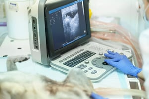
- Abdominal ultrasound to evaluate intestinal wall thickness, lymphadenopathy and other structural abnormalities; a normal result does not exclude CIE
While not GI-specific, basal cortisol testing may also be indicated in dogs with chronic GI signs to rule out hypoadrenocorticism. Following this initial work-up, the next step may include a structured diet or treatment trial, further diagnostic investigation, or both, depending on the clinical picture.
Diet or treatment trials and empirical management
In practice, not every dog proceeds directly to biopsy. In clinically stable patients, a structured diet or treatment trial may be considered before more invasive investigation, particularly where signs are not severe or where owners are reluctant to pursue endoscopy or surgery at the outset. This is also reflected in recent ACVIM guidance, which suggests that ideally at least three properly designed dietary treatment trials should be carried out before invasive procedures such as endoscopy are pursued (Heilman et al., 2026).
This is one reason CIE is often classified retrospectively according to response to treatment. Food-responsive enteropathy (FRE) is the most common subtype, with one study suggesting that more than half of dogs with CIE fall into this category (Jergens & Heilmann, 2022). Dietary management remains a cornerstone of treatment, with response rates as high as 88% (Jugan, 2020). A marked improvement or complete resolution of clinical signs following a trial with a novel or hydrolysed diet supports a diagnosis of FRE.
While this staged approach is often appropriate, one of the challenges in practice is determining whether intestinal inflammation is actually present and how it is changing over time, for example in response to diet trial. In this context, faecal biomarkers may provide additional objective data to support assessment and monitoring.
What is faecal calprotectin?
Faecal calprotectin is a sensitive biomarker of gastrointestinal inflammation (Dabritz, 2014; Heilman, 2008; Jukic, 2021). During gastrointestinal inflammation, the innate and adaptive immune responses are stimulated, resulting in an influx of phagocytic cells and the release of inflammatory cytokines. These chemoattract neutrophils, triggering a cascade of events leading to neutrophil disintegration (Figure 1) (Dabritz, 2014; Jukic, 2021; Heilman, 2012).

Figure 1. Mechanism of faecal calprotectin release during gastrointestinal inflammation. 1. Immune responses are stimulated. 2. Release of inflammatory cytokines. 3. Neutrophil disintegration results in calprotectin release into the gut lumen.
Calprotectin makes up around 60 percent of the neutrophil’s cytosol (fluid within a cell’s cytoplasm). Neutrophil disintegration therefore results in calprotectin being released into the gut lumen, where it enters the faecal stream, and can then be measured.
Importantly, calprotectin is resistant to degradation by faecal bacteria and its concentration in faeces therefore correlates with the degree of neutrophilic infiltration in the gastrointestinal mucosa.
Faecal calprotectin at point-of-care
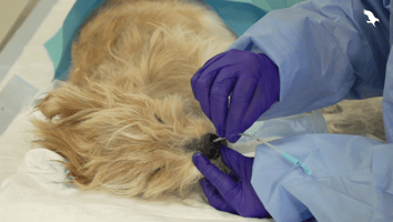
Patient-side faecal calprotectin testing offers a practical way to assess intestinal inflammation in dogs with chronic GI signs. As a non-invasive test performed on a faecal sample, it can be incorporated into the diagnostic work-up without the need for sedation, anaesthesia, or surgery.
In practice, point-of-care faecal calprotectin may be particularly useful in dogs where:
- chronic GI signs are present, but the degree of underlying inflammation is unclear
- owners are reluctant to pursue invasive diagnostics
- financial constraints limit the extent of the initial work-up
- support is needed for decision-making around diet or treatment trials
- support is needed for owner communication
One commercially available patient-side faecal calprotectin test has shown good diagnostic performance in dogs with chronic enteropathy, with specificity of 96 percent and sensitivity of 94 percent at a threshold of 3 mg/kg (Carus Animal Health, 2025).
Used alongside the wider clinical picture, faecal calprotectin can provide objective information about intestinal inflammation. It does not replace histopathology or other investigations, but may provide useful additional information when deciding on the next step.
Role of histopathology
Histopathology remains the gold standard for the definitive diagnosis of canine chronic inflammatory enteropathy and for ruling out other important differentials, including neoplasia. Intestinal biopsy samples may be obtained endoscopically, allowing collection of mucosal biopsies from accessible regions of the GI tract, or surgically, where full-thickness samples can be collected if indicated. In dogs with chronic GI signs, intestinal biopsy can provide direct information about the type, distribution and severity of mucosal inflammation, and may help distinguish inflammatory disease from other infiltrative or structural pathology.
Histopathology is particularly important where:
- Clinical signs are severe, progressive, or poorly responsive to initial management
- Important differentials such as neoplasia remain a concern
- A definitive diagnosis is needed to guide prognosis and longer-term treatment planning
Histopathology may be performed as part of the initial diagnostic investigation. However, a treatment or dietary trial is often considered first, following the ACVIM consensus guidelines,3 with biopsy pursued only if response is poor or clinical signs are severe.
It is important to note that while histopathology of intestinal biopsies is required for definitive diagnosis, the severity of clinical signs does not always reliably predict the extent of histological lesions (Heilman, 2014).
Take-home points
- Diagnosis of canine chronic inflammatory enteropathy requires a structured diagnostic plan
- Initial investigation should include history, clinical examination, routine laboratory testing, GI-specific tests and imaging where indicated
- In clinically stable dogs, a diet or treatment trial may be appropriate before more invasive investigation
- Histopathology remains the gold standard for definitive diagnosis and for ruling out important differentials such as neoplasia
- Faecal calprotectin can provide objective information about intestinal inflammation, but should be interpreted alongside the wider clinical picture