Placement of a naso-oesophageal (N-O) feeding tube or naso-gastric (N-G) feeding tube is well tolerated and extremely helpful in small animal practice. In the step-by-step video below, we explain how to place a naso-oesophageal or naso-gastric feeding tubes in dogs and cats.
Step by step placement of a naso-oesophageal feeding tube
The video below shows the placement of a naso-oesophageal feeding tube.
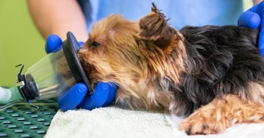
Step 1: Prepare the patient and tube
Anaesthetic and sedation are not usually needed. Numb the patient's nostril with local anaesthetic (proxymetacaine or lidocaine). Take the feeding tube (91cm long; 5Fr for cats, 5-8Fr for dogs) and measure the length, marking it so that you know how far in to advance the tube. Lubricate the end of the tube fully.
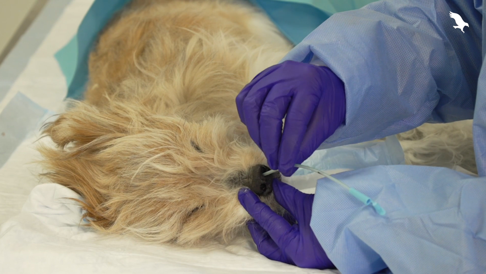
Step 2: Inserting the tube
Guide the tube into the nostril, directing it medially and centrally. Advancing it, it should pass through the ventral meatus without meeting resistance, all the way to where the premeasured mark is on the tube. If resistance is felt at the point of the median septum (2-3cm in), push the external nares dorsally ('pig nose') to open up the ventral meatus.
Step 3: Checking for correct placement
The following ways are useful to check for correct placement of an naso-oesophageal tube:
- You may see the patient swallow as the tube moves past the throat and into the oesophagus.
- Using an empty syringe, check for negative pressure in the tube - if there is no negative pressure, you may be in the trachea, while fluid may be from gastric juices.
- Attaching a capnograph to the tube can be a good way to check for incorrect tube placement. Any end-tidal CO2 suggests inadvertent airway positioning.
- A small amount of sterile saline dripped into the tube should elicit a cough if the tube is in the trachea.
- A lateral radiograph can help to visually confirm tube placement if the patient is amenable or sedated.
Step 4: Securing the tube
Once you've confirmed the correct placement of the naso-oesophageal feeding tube, secure it to the patient using a Chinese finger trap suture. You may need to anchor the other end of the tube, either to the patient's face or further down their body to stop it flapping around and bothering them.
Indications for feeding tubes in small animals
Feeding tubes are placed to manage conditions that cause anorexia (either partial or complete) that's expected to last more than 3 days. Naso-oesophageal feeding tubes and naso-gastric feeding tubes are used when the support is expected to be required only short-term (3-7 days). Because N-O and N-G tubes don't require sedation to place, they may also be used temporarily where the patient isn't stable enough for a longer-term oesophagostomy tube to be placed.
Common indications for feeding tubes include:
- Acute pancreatitis
- Gastrointestinal disease (eg parvovirus, inflammatory bowel disease causing inappetence)
- Liver disease (especially cholangiohepatitis in cats)
- Trauma (especially to the face) such as dog fights or road traffic accidents
- Acute kidney injury
- Any catabolic state where their intake (or absorption) isn't keeping up with their needs (eg severe burns, protein-losing enteropathy, septic peritonitis)
The main contraindication to N-O or N-G tubes is pets with nasal disease, who may do better with an oesophagostomy tube. Other contraindications include:
- Oesophageal disease (such as oesophagitis or megaoesophagus)
- Where there is a high risk of aspiration because the animal is comatose or laterally recumbent
- Uncontrolled persistent vomiting (because the tube may be expelled or misdirected)
- Where a pure liquid diet is not suitable (and therefore a larger bore tube that can take gruel diets is required)
In which case, other types of feeding support may be preferred.
Managing pets with a feeding tube
Once pets have the naso-oesophageal feeding tube in place, they need to be fed a suitable diet. For naso-oesophageal tubes, this needs to be a complete liquid diet, and (apart from the first few days, where it's introduced gradually) should meet their Resting Energy Requirement (RER), which should be recalculated daily to ensure the needs are met.
The tube needs flushing before and after each feeding to ensure it doesn't block and the placement is still correct.
In many cases, patients with N-O feeding tubes remain hospitalised, where they can receive other necessary medications and complications can be managed. However, in a recent review (Dumont 2023) it was shown that naso-oesophageal tubes may be able to be managed at home - while complications were common, they were rarely serious.
Removing the feeding tube
The feeding tube is removed by cutting the suture then pulling the tube gently out. This is usually tolerated in conscious animals.
References:
Dumont