In general practice the term interdigital cyst is commonly used to describe a range of conditions affecting the feet. Some use the term to describe any swelling and inflammation in the interdigital webs, others use it for inflammation of any part of the foot. However, a cyst is defined as a well-circumscribed epithelium-lined cavity filled with solid material or with fluid. In the skin these occur as solid or fluctuant masses which may or may not be lined with epithelium of the adnexal structures. It is important to distinguish these from other forms of pododermatitis, the term used to describe inflammation of the foot involving the interdigital skin, claw beds and claws.
This article will focus on raised swellings in skin between the digits rather than the more diffuse inflammation affecting the feet. So, before we can answer the question of “are they curable?”, we need to ask “why do these occur?”
Primary causes
Probably the most common primary causes include allergies and foreign bodies. Pedal pruritus is common in dogs with atopic dermatitis. Persistent licking pushes the hairs into the dermis and subcutis resulting in inflammation, hair follicle rupture and free keratin, all of which can result in cyst formation.
Grass awns and other penetrating foreign bodies cause a soft tissue reaction resulting in an “interdigital granuloma”. Primary bacterial or fungal infections are mainly introduced through penetrating wounds resulting in pyogranulomas, with or without draining sinuses. Adverse drug reactions, sterile pyogranuloma and hormonal and metabolic causes are uncommon to rare.
Before the introduction of the isoxazolines on the veterinary market, Demodex mites were often seen as a cause of interdigital cysts. Since many practices now use afoxolaner, fluralaner, lotilaner or sarolaner as routine prophylactic flea and tick treatments, interdigital cysts associated with Demodex mites are uncommon. However, it is important to include demodicosis as a potential differential diagnosis during investigations.
Orthopaedic problems such as elbow dysplasia, osteoarthritis, hip dysplasia and other orthopaedic problems that may result in an abnormal gait contribute to the disease process. Some dogs are born with congenital deformities such as splayed feet. Some dogs with these problems walk on haired skin surrounding the digital pads. The highly keratinised tissue of the pads is capable of withstanding friction and protects the foot. However, when dogs walk on the haired skin the hairs start to become embedded in deeper tissue initiating a foreign body reaction. In early stages comedones are visible and with repeated friction the hairs start to form cystic follicles which progress to formation of large cysts. Follicular cysts on the ventral aspect of the paws form a sinus tract leading to the dorsal aspect of the foot and appear as a nodule/swelling. The swelling often ruptures, ulcerates and drains serosanginous fluid.
There is a subset of dogs where an underlying or predisposing factor cannot be identified even after extensive investigation. In one study where no underlying disease was identified, dogs with recurring interdigital cysts responded to immunomodulatory treatment with either glucocorticoids or ciclosporin (Breathnach et al., 2005). Histopathological findings in these dogs included lymphocytic-plasmacytic infiltrate and suspected immune-mediated reaction.
Predisposing factors
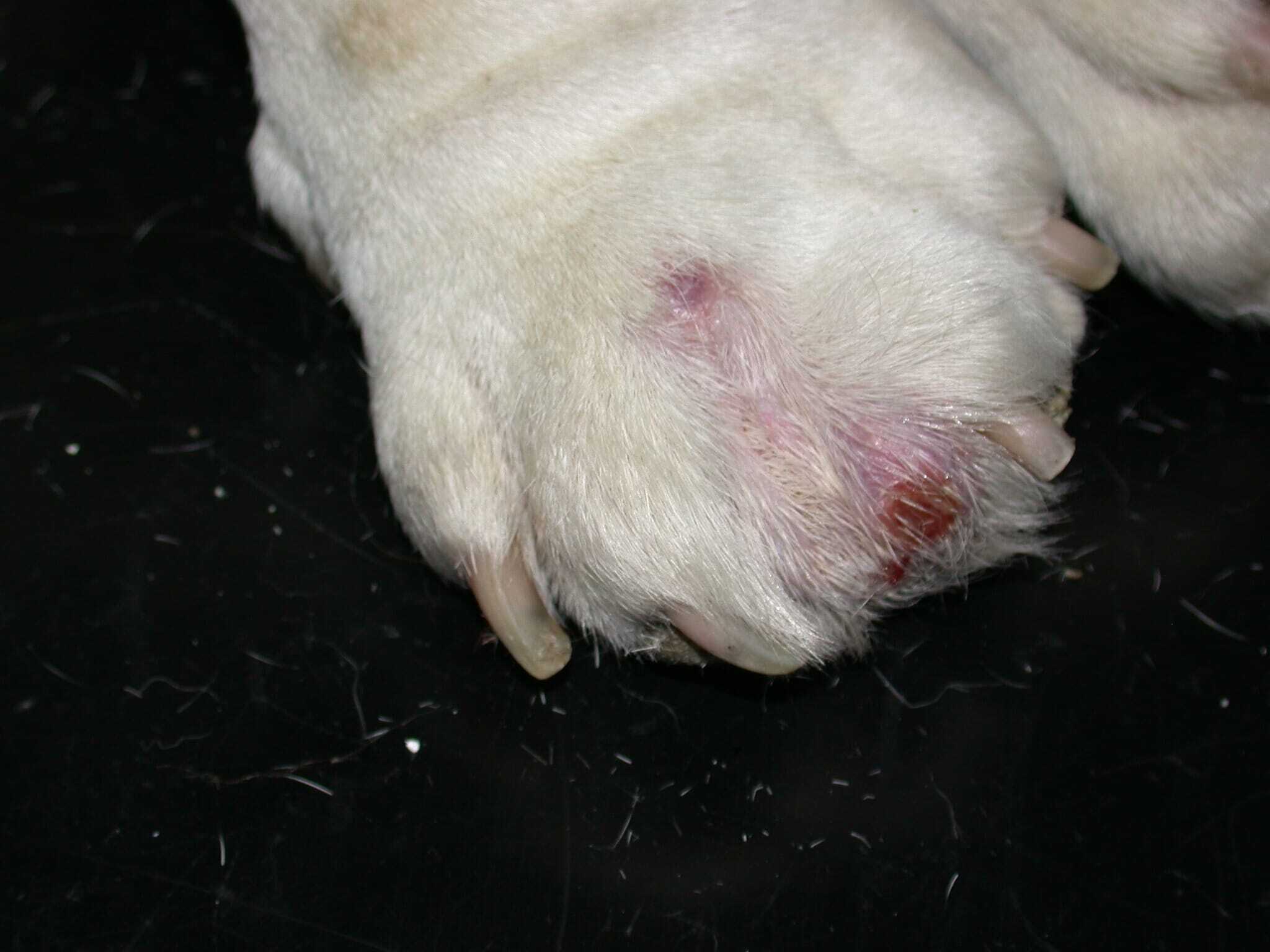
The length and thickness of the hair can be a predisposing factor. Short hair, especially thick short hair, is more likely to penetrate the soft tissue due to persistent licking or friction on haired skin surrounding the pads caused by abnormal gait or conformation. Short-haired dogs such as Boxers, Bulldogs, Mastiffs, Great Danes and Bull Terriers are predisposed to interdigital cysts. Obesity, spinal disease and conformational and gait abnormalities contribute to formation of false pads and in the long term progress into cystic lesions. Poor conformation, such as widely splayed toes ( Figure 1A and 1B), may favour traumatic injury to interdigital webs.


Perpetuating factors
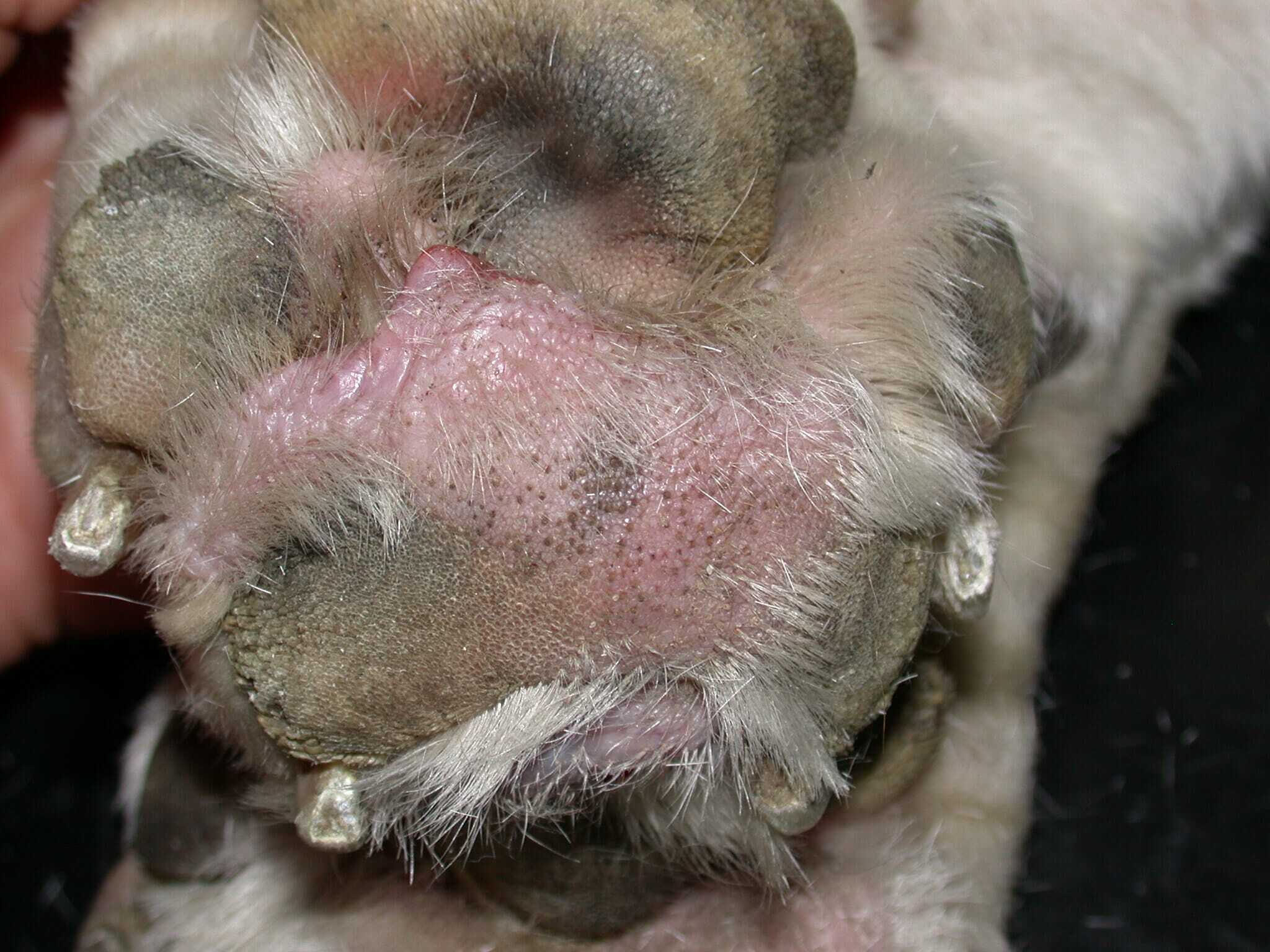
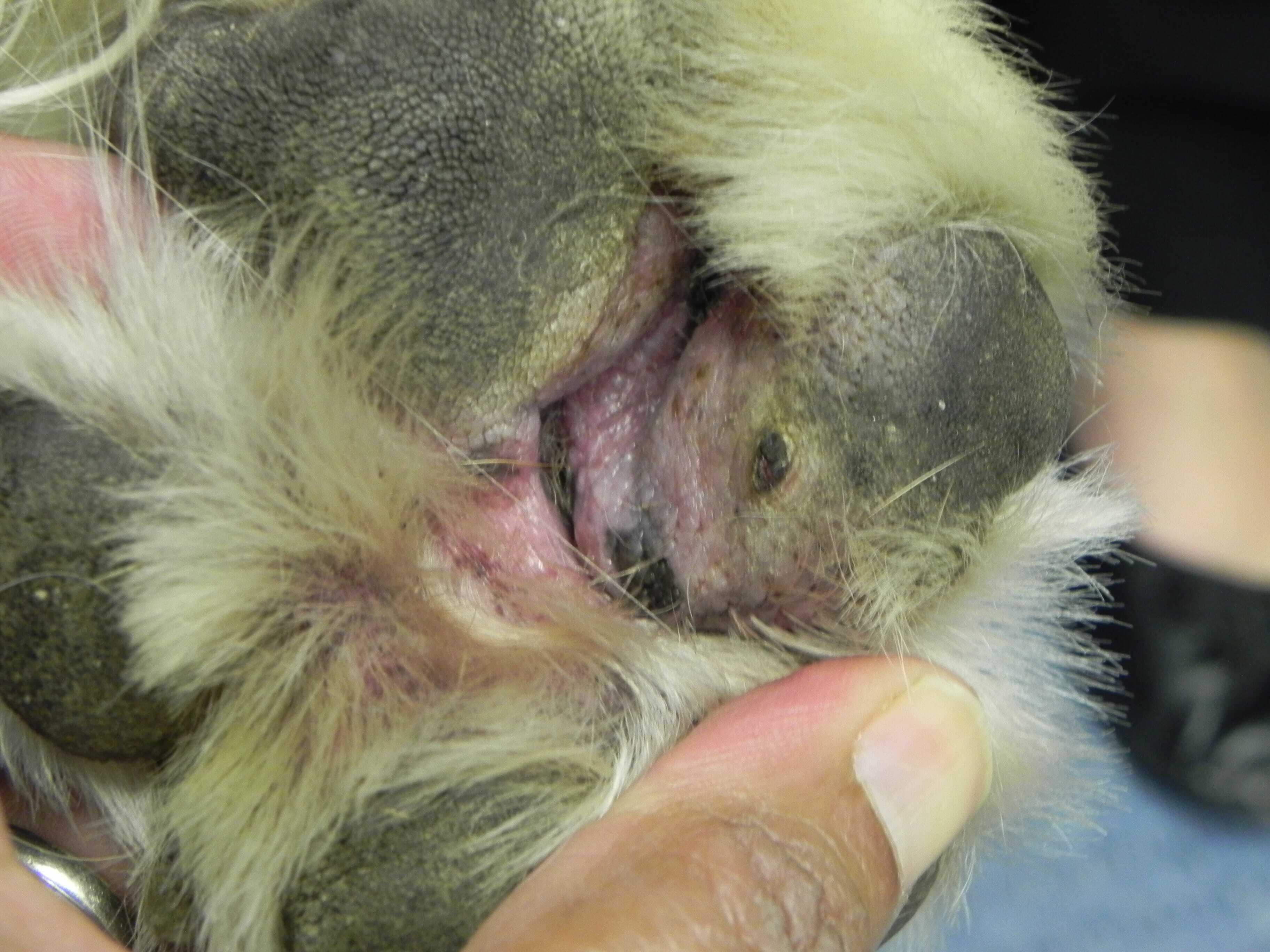
Ongoing and persistent soft tissue proliferation results in formation of “false pads” (also sometimes referred to as pseudo pads) or conjoined pads ( Figure 2A and 2B). This is a common perpetuating factor in brachycephalic breeds which tend to bear more weight on the anterior aspect of the body. Hyperkeratosis and deep crevices ( Figure 3) which harbour debris and ingrowing hairs are contributory factors. Previous episodes of furunculosis may trigger ongoing disease because of free hair shafts and free keratin in the dermis.
Secondary infections
Bacterial infections exacerbate the signs resulting in an acute event where the cyst ruptures and releases purulent exudate. Staphylococcus pseudintermedius is most commonly implicated; however, mixed infections with Streptococcus and Gram-negative species such as E. coli, Pseudomonas spp. and Proteus spp. may be involved. Malassezia and other fungal organisms may also play a role in perpetuating the lesions.
Summary
So, in summary, a cure for a condition involving multiple factors is likely to be challenging and often requires long-term management in most dogs with interdigital cysts. It is best to inform the owner at the outset of the multifactorial nature of the condition. It needs a multimodal approach which requires dermatological therapies, orthopaedic assessment and/or soft tissue surgery to provide a successful outcome.
The dermatological treatment for these recurrent cysts includes four to eight weeks (sometimes longer) of antibiotic therapy based on culture and sensitivity for secondary infections. The management of allergic skin disease is essential. Weight loss in obese dogs is advised. Changing areas where the dogs are walked and shorter, more frequent walks may be better than one long walk. Management in each case should be individualised. The soft tissue can be ablated with a CO2 laser in dogs with false pads or excess granulation tissue. In more severe cases, podoplasty, a radical surgical excision of interdigital skin, may represent a last resort form of approach if medical management fails. But remember, these are measures to improve welfare and are not a cure.
References (click to expand)
| Breathnach, R. M., Baker, K. P., Quinn, P. J., McGeady, T. A., Aherne, C. M. and Jones, B. R. | 2005 | Clinical, immunological and histopathological findings in a subpopulation of dogs with pododermatitis. Veterinary Dermatology, 16, 364-372 |
| Breathnach, R. M., Fanning, S., Mulcahy, G., Bassett, H. F., Jones, B. R. and Daly, P. | 2006 | Evaluation of Th1-like, Th2-like and immunomodulatory cytokine mRNA expression in the skin of dogs with immunomodulatory-responsive lymphocytic-plasmacytic pododermatitis. Veterinary Dermatology, 17, 313-321 |
| Dulcos, D. D. | 2013 | Canine pododermatitis. Veterinary Clinics of North America: Small Animal Practice, 43, 57-87 |
| Dulcos, D. D., Hargis, A. M. and Hanley, P. W. | 2008 | Pathogenesis of canine interdigital palmar and plantar comedones and follicular cysts, and their response to laser surgery. Veterinary Dermatology, 19, 134-141 |
| Seppänen, R. T., Kaimio, M., Schildt, K. J., Lilja‐Maula, L., Hyytiäinen, H. K., Mölsä, S., Morelius, M., Rajamäki, M. M., Lappalainen, A. K. and Rantala, M. | 2019 | Skin and ear health in a group of English bulldogs in Finland – a descriptive study with special reference to owner perceptions. Veterinary Dermatology, 30, 307–e85 |