Malassezia dermatitis (also referred to as Malassezia overgrowth syndrome) and otitis may be more prevalent than recognised. Malassezia infections are common in allergic dogs but there is little information in cats. A small number of these yeast organisms are found on healthy cats; however, a breakdown in the epidermal barrier and changes in the microbiome lead to an overgrowth of the organism. Malassezia pachydermatis is the organism most associated with infections, but other species such as M. sympodialis, M. nana, M. furfur and M. globosa (Bond et al., 1996, 1997; Crespo et al., 1999, 2000) have been implicated in infections.
Risk factors
The factors that contribute to the shift in Malassezia population in cats are:
Genetics
Some breeds may have a higher carriage rate of Malassezia on the skin and ears. Healthy Devon Rex cats were shown to have higher carriage of Malassezia on the skin which may be one of the reasons why they are predisposed to seborrhoeic dermatitis (Bond et al., 2008). A breed predisposition is recognised in Sphynx cats and in both Devon and Cornish Rex cats (Ahman et al., 2007; Ahman and Bergstrom, 2009).
Allergies
Sensitivities to environmental and dietary allergens have been associated with an overgrowth of Malassezia (Ordeix et al., 2007), but it is important to note that not all allergic cats develop Malassezia dermatitis.
Immune dysregulation and systemic disease
In one report Malassezia was isolated more frequently from retroviral positive cats compared to those that were negative (Sierra et al., 2000), which suggests that overgrowth may be linked to an underlying immune dysregulation. Immunosuppression associated with anti-inflammatory drugs, like glucocorticoids and immunomodulators such as ciclosporin and chlorambucil, could be a contributing factor to population changes during the management of the primary diseases. In some cases, Malassezia overgrowth may indicate systemic disease (eg inflammatory bowel disease, hyperadrenocorticism, diabetes mellitus, hyperthyroidism) and/or paraneoplastic syndromes (eg exfoliative dermatitis associated with a thymoma, paraneoplastic alopecia, systemic neoplasia) (Forster-Van Hijfte et al., 1997; Godfrey, 1998; Perrins et al., 2007). If large numbers of organisms are found during a case work-up, a detailed history, clinical signs, laboratory tests and imaging modalities relating to the underlying disease are indicated to reach a diagnosis and prognosis. Note that some of these conditions have a poor to guarded prognosis.
Clinical signs
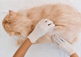
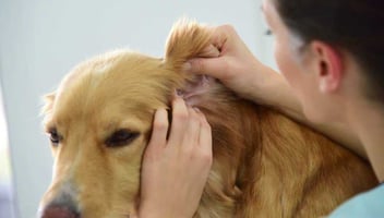
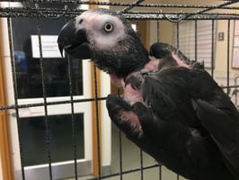
The clinical signs are variable and may well depend on the underlying condition, and there is no particular distribution pattern. Not all cases of Malassezia infections are pruritic in the initial stages, but can become so with time. The lesions include dry ( Figure 1) or greasy scale, erythema, follicular casts, brown exudate ( Figure 2), alopecia and hyperpigmentation. Cats with Malassezia otitis have brown to black ceruminous exudate in the ear canals ( Figure 3), stenosis, erythema and self-inflicted lesions on the preauricular skin. Accumulation of brown to black exudate on the claw folds or in the claws are seen in some cases ( Figure 4) as well as on the feet ( Figure 5). Some of these cats are presented with a history of claw or feet biting and licking. Predisposed breeds such as the Devon Rex present with seborrhoeic dermatitis.


Diagnosis
The diagnosis is based on history, clinical signs, demonstration of the organisms on cytology and response to antifungal treatment. Culture for Malassezia on the skin is not generally done; however, their presence is often reported on ear swabs.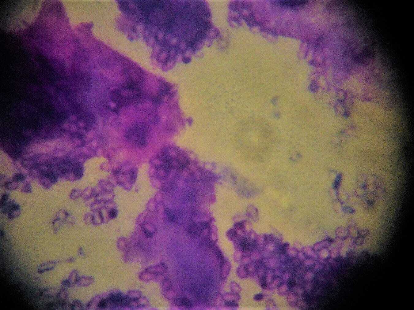
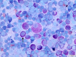
Cytology
Malassezia organisms are footprint to ovoid in shape, and the quickest way to identify them is using tape strips on the skin ( Figure 6) and/or swabs from ears, stained in Diff Quik.
Response to treatment
To ascertain the role of Malassezia in the severity of lesions and/or their contribution in increasing the level of pruritus, it is best to treat. A reduction in pruritus or lesion scores supports their role in the disease.
Culture
M. pachydermatis is readily cultured using standard mycological techniques as it is non-lipid dependent, but other Malassezia spp. require a lipid-enriched culture medium to grow on.
Histopathology
Skin biopsies may show large numbers of Malassezia in the stratum corneum. The presence of very large numbers of organisms in the absence of an allergy could be suggestive of immune dysregulation and/or systemic disease.
Treatment
Treatment should be individualised on a case-by-case basis. It will depend on the extent and severity of the lesions, the underlying disease, owner compliance and the temperament of the cat. Topical treatment is most useful for cats with otitis. Ear preparations containing miconazole, clotrimazole and nystatin are licensed for cats in the UK. Shampoo treatment using miconazole/chlorhexidine shampoo is useful ( Figure 7), but cats are not the easiest animals to bathe. Mousse products containing climbazole/chlorhexidine may be useful for some individuals.


Systemic antifungals are indicated if topical treatment is ineffective or impractical. Itraconazole at 5mg/kg once daily orally, as a pulse treatment of seven days on and seven days off, can be used. A marked improvement in clinical signs with a reduction in pruritus and inflammation has been demonstrated in studies (Bensignor, 2010; Ahman et al., 2007). Generally, two cycles are sufficient to assess the response to treatment.
Summary
Given that the cutaneous microbiome can be affected by a wide range of skin diseases, overgrowth of Malassezia can occur with any of them. Unless one specifically looks for their presence on the skin in cats with pruritus and/or dermatitis, they can be missed, which results in difficult to manage primary diseases and ongoing client dissatisfaction. Malassezia dermatitis and otitis are easy to diagnose and treat, resulting in better and more successful management of primary diseases.
_OL_CourseThumb.jpg?width=500&height=300&name=IVE_GPCert(Derm)_OL_CourseThumb.jpg)
Dermatology
ISVPS General Practitioner Certificate (GPCert)
Postgraduate Certificate (PgC)
 May 2026
 Online
References (click to expand)
| Ahman, S. E. and Bergstrom, K. E. | 2009 | Cutaneous carriage of Malassezia species in healthy cats and seborrheic Sphynx cats and a comparison to carriage in Devon Rex cats. Journal of Feline Medicine and Surgery, 11, 970-976 |
| Ahman, S., Perrins, N. and Bond, R. | 2007 | Treatment of Malassezia pachydermatis‐associated seborrhoeic dermatitis in Devon Rex cats with itraconazole – a pilot study. Veterinary Dermatology, 18, 171-174 |
| Bensignor, E. | 2010 | Treatment of Malassezia overgrowth with itraconazole in 15 cats. Veterinary Record, 167, 1011-1012 |
| Bond, R., Anthony, R. M., Dodd, M. and Lloyd, D. H. | 1996 | Isolation of Malassezia sympodialis from feline skin. Journal of Medical and Veterinary Mycology, 34, 145-147 |
| Bond, R., Howell, S. A., Haywood, P. J., Lloyd, D. H. | 1997 | Isolation of Malassezia sympodialis and Malassezia globosa from healthy pet cats. Veterinary Record, 141, 200-201 |
| Bond, R., Stevens, K., Perrins, N. and Ahman, S. | 2008 | Carriage of Malassezia spp. yeasts in Cornish Rex, Devon Rex and Domestic short-haired cats: a cross-sectional survey. Veterinary Dermatology, 19, 299-304 |
| Crespo, M. J., Abarca, M. L. and Cabanes, F. J. | 1999 | Isolation of M. furfur from a cat. Journal of Clinical Microbiology, 37, 1573-1574 |
| Crespo, M. J., Abarca, M. L. and Cabanes, F. J. | 2000 | Otitis externa associated with M. sympodialis in two cats. Journal of Clinical Microbiology, 38, 1263-1266 |
| Forster-Van Hijfte, M. A., Curtis, C. F. and White, R. N. | 1997 | Resolution of exfoliative dermatitis and M. pachydermatis overgrowth in a cat after surgical thymoma resection. Journal of Small Animal Practice 1997, 38, 451-454 |
| Godfrey, D. R. | 1998 | A case of feline paraneoplastic alopecia with secondary Malassezia-associated dermatitis. Journal of Small Animal Practice, 39, 394-396 |
| Ordeix, L., Galeotti, F., Scarampella, F., Dedola, C., Bardagí, M., Romano, E. and Fondati, A. | 2007 | Malassezia spp. overgrowth in allergic cats. Veterinary Dermatology, 18, 316-323 |
| Perrins, N., Gaudiano, F. and Bond, R. | 2007 | Carriage of Malassezia spp. yeasts in cats with diabetes mellitus, hyperthyroidism and neoplasia. Medical mycology, 45, 541-546 |
| Sierra, P., Guillot, J., Jacob, H., Bussiéras, S. and Chermette, R. | 2000 | Fungal flora on cutaneous and mucosal surfaces of cats infected with feline immunodeficiency virus or feline leukemia virus. American journal of veterinary research, 61, 158-161 |