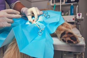
A surgical site infection is never ideal, but in oncology cases it can feel particularly frustrating. These are often patients where the surgery has already required a lot of planning, the owner may be anxious, and the next step — whether that’s histopathology, staging, chemotherapy, radiotherapy or simply getting the dog back to normal — depends on the wound healing as expected.
Of course, experienced RVNs don’t need an article to tell them that clean clippers, good skin prep and keeping patients warm are useful things. But oncology surgery does have a few extra quirks that are worth thinking through. Mass removals can mean wide margins, long incisions, awkward locations, ulcerated tissue, drains, dressings, reconstruction, longer anaesthetic times and older patients with comorbidities. So, while preventing infections at the surgical site still comes back to familiar nursing principles, the stakes — and the practical details — can be a little different.
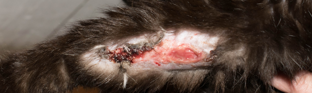
First, a quick reminder: what counts as a surgical site infection?
Most nurses will recognise an infected wound when they see one. Purulent discharge is rarely subtle, and the “that doesn’t look right” feeling at a postop check is often well worth listening to.
However, when we’re talking about the evidence, it helps to be clear about definitions. Surgical site infections are usually grouped as superficial incisional, deep incisional, or organ/space infections, and many studies use a 30-day postoperative follow-up period. Some include only purulent discharge, abscessation or fistulation; others also include combinations of redness, swelling, heat, pain, serous discharge and dehiscence (Eugster et al., 2004; Espinel-Rupérez et al., 2019).
That matters because SSI rates vary depending on how hard you look for them and what you count. A practice that only records the cases that come back with obvious discharge will get a very different number from one that actively follows up every patient at 7 and 30 days. This is one reason studies report slightly different figures — and why good nursing records and audit systems are so useful.
What's different about oncological surgery?
The phrase “mass removal” can hide a lot. A small, freely movable, non-ulcerated lump on the lateral thorax is one thing. A large mammary mass involving the inguinal region, an ulcerated mast cell tumour on the distal limb is another. The nursing plan for these cases may include the same basic principles, but the emphasis changes.
In oncology surgery, SSI risk may be affected by:
- larger surgical fields
- longer prep times
- longer anaesthetic and surgical times
- ulcerated, inflamed or contaminated tumour surfaces
- increased dead space and the need for drains
- more tension across the wound
- reconstructed wounds or flaps
- patient factors such as age, body condition, and comorbidities
- the consequences of delayed healing, especially where adjuvant treatment is planned
Several veterinary studies have examined risk factors for SSIs. In a large multicentre study of clean surgical procedures in dogs, surgery time was the only variable associated with increased SSI risk (Stetter et al., 2021). Another study found SSI in 8.7% of dogs, with risk factors including surgery lasting over 60 minutes, preoperative hyperglycaemia, steroidal anti-inflammatory use, urinary catheterisation and lack of Elizabethan collar use (Espinel-Rupérez et al., 2019).
That doesn’t mean oncology surgery is automatically high-risk, and many of these surgeries are clean. But thinking about the small ways to reduce risk over the entire perioperative period can make a big difference to individual cases.
Before surgery
Understanding the surgical plan
For oncology patients, it’s worth taking a moment to check exactly what is being removed, where it is, and whether the plan might change once the patient is clipped, positioned or draped.
Useful questions include:
- Are there multiple masses?
- Has the mass been measured/photographed?
- Is the mass ulcerated, inflamed, necrotic or discharging?
- Is wide excision planned?
- Might the surgeon need to extend the incision?
- Is there a chance of a drain, dressing, tie-over dressing, reconstruction, or staged procedure?
- Are samples needed for histopathology, culture or margin assessment?
- Is the patient likely to need chemotherapy or radiotherapy once the wound has healed?
This is not about RVNs second-guessing the surgeon. It is about making sure the nursing team is prepared. For example, if the case says “mammary mass removal”, the practical nursing implications may be very different depending on whether this is a lumpectomy, regional mastectomy or chain mastectomy. In one study of dogs undergoing mastectomy for mammary gland tumours, 16.9% of surgeries were followed by complications, and more extensive procedures were associated with higher complication risk (Evans et al., 2021). That has obvious implications for preparation, analgesia, recovery monitoring and owner instructions.
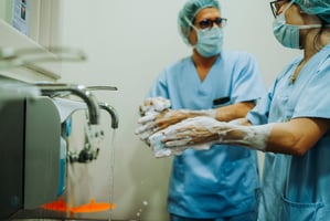
Clip widely, but kindly
Skin preparation is familiar territory, but oncology cases can make the “how wide do we clip?” question more interesting than usual.
Wide margins, possible incision extension, drains and dressings all need to be considered before the first clipper line is made. A clip that is perfectly adequate for the mass itself may not be adequate if the surgeon needs a wider approach or a drain exit site. (Nobody wants to be trying to extend a clip around a patient who is already positioned, partially prepped and surrounded by sterile kit!)
At the same time, skin trauma matters. Clipper rash, repeated passes over thin skin, or aggressive clipping around inflamed tissue may all make the postoperative wound less happy than it needed to be. There is a balance between generous preparation and unnecessary trauma.
Where masses are ulcerated or contaminated, it may be sensible to think about the direction of cleaning and whether separate prep materials are needed, so debris is not simply moved from the tumour surface across the future surgical field.
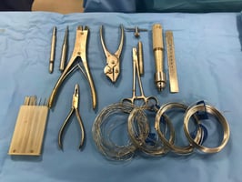
Theatre set-up
Oncology procedures can run longer or become more complex than expected, so theatre set-up deserves a little more thought.
Depending on the case, this might include:
- extra packs of sterile gloves and gowns
- extra packs of drapes
- additional instruments or a second instrument pack - eg if switching kits to reduce seeding of tumour cells, see below
- lavage
- suction
- haemostatic materials and dressings
- drains
- stuff for histology, eg sample pots and labels and margin-marking materials
- warming equipment
For nurses, this is one of the areas where preparation pays off. Having the right kit ready before the patient is induced is rarely exciting, but it is much better than trying to find the right dressing while everyone is scrubbed, the wound is open and the anaesthetic clock is ticking.
And yes, you can always call for more kit, but in Eugster et al.’s prospective study, increasing numbers of people in the operating room were associated with SSI in one of their models (Eugster et al., 2004). That does not mean every observer causes an infection by existing, but it is a helpful reminder that theatre traffic is not just distracting, it can add time and additional risk.
A note on oncology-specific theatre protocols
There is another oncology-specific consideration here: tumour seeding.
This is separate from SSI, but relevant to theatre planning. A recent survey of veterinary surgeons found that most respondents reported changing gloves and instruments during oncological surgery to reduce the risk of tumour seeding, although wound-edge protection was less commonly reported (Orjefelt et al., 2025). There is not yet clear veterinary evidence that retained neoplastic cells on instruments or gloves cause recurrence, so this is an area where protocols vary. Still, it is increasingly discussed, especially in referral and surgical oncology settings.
For RVNs, the practical point is to know the surgeon’s plan. If your team uses separate “dirty” and “clean” instruments, changes gloves after tumour handling, separates areas on the instrument table, or uses wound protection, those steps need to be anticipated.
During surgery: time, temperature, antibiotics and theatre discipline
Perioperative antibiotics
Antimicrobials are often discussed in relation to SSI prevention, but the evidence is not as simple as “give antibiotics and the wound will be fine”.
Some procedures will need perioperative antibiosis, especially where they are clean-contaminated, contaminated, prolonged, involve implants, or where patient factors increase concern. Others may not. The decision is the veterinary surgeon’s, but RVNs are often the people who make sure the plan actually happens at the right time.
That means:
- checking whether antibiotics have been prescribed
- giving them at the correct time, according to practice protocol
- recording the time accurately
- prompting re-dosing if the procedure is prolonged and the protocol requires it
- recording any missed, delayed or repeated doses clearly
- supporting culture and sensitivity if infection later develops
In the Stetter et al. study, perioperative antimicrobial prophylaxis was not associated with SSI development in clean procedures, whereas surgery time was (Stetter et al., 2021). Espinel-Rupérez et al. also found no benefit to continuing chemoprophylaxis beyond 24 hours postoperatively in their cohort (Espinel-Rupérez et al., 2019). That does not mean antibiotics are never useful, but they aren't a magic bullet.
The nursing role here is very much an antimicrobial stewardship role: accurate timing, accurate recording, and gentle prompting when needed. Especially in longer oncology procedures, this can make a real difference to whether the intended protocol is followed.
Anaesthetic monitoring
One of the interesting things about SSI prevention is how often the apparently “surgical” risk factors are actually shared across the whole theatre team.
Duration of anaesthesia and surgery have been associated with postoperative wound infection in several studies (Beal et al., 2000; Nicholson et al., 2002; Eugster et al., 2004; Stetter et al., 2021). In clean-contaminated procedures, Nicholson et al. found that animals developing wound infections had significantly longer surgery and anaesthesia times (Nicholson et al., 2002). Stetter et al. found that, in clean procedures, surgeries over 120 minutes had higher odds of SSI than procedures under 30 minutes (Stetter et al., 2021).
Of course, nurses cannot make a difficult tumour resection suddenly take 20 minutes. Nor should speed ever come at the expense of good surgery. But you can influence the time around the surgery: smooth induction, efficient clipping and prep, appropriate theatre set-up, good communication and avoiding delays once the patient is anaesthetised.
Temperature is another area where the picture is nuanced. Beal et al. did not find mild perioperative hypothermia to be a significant risk factor for wound infection in clean wounds, but duration of anaesthesia was significant (Beal et al., 2000). Other studies and clinical guidance continue to emphasise normothermia for broader perioperative reasons, including perfusion, coagulation, recovery quality and patient comfort. So, even where the SSI link is not straightforward, temperature management remains good nursing.
In potentially lengthy oncological surgeries, that means:
- planning warming before the patient is cold
- monitoring temperature regularly
- using active warming appropriately
- minimising unnecessary wetting during prep
- avoiding long waits between induction, prep and incision
- monitoring perfusion, blood pressure and oxygenation, and flagging issues early
Theatre discipline during longer oncology cases
Longer procedures can make small lapses more likely. People get tired. Gloves become contaminated. Drapes get damp. Someone opens the theatre door to ask a quick question. The patient needs repositioning. The sample pot is not labelled. The suction tubing is not where anyone thought it was. Remember, these problems may be little, but they're cumulative.
During oncology surgery, RVNs may be involved in:
- maintaining a sterile field during a long procedure
- watching for breaks in asepsis
- keeping theatre traffic sensible
- helping with glove or instrument changes if used
- separating tumour-handling instruments from closure instruments where required
- managing samples without contaminating the field
- ensuring dressings, drains or lavage are ready when needed
Not to mention monitoring the anaesthetic!
This is advanced nursing in a very practical sense, and where the experienced RVN can really make a difference to patient safety. Training in theatre management can make a big difference in these cases.
After surgery: the wound still needs nursing
Recovery period
SSI prevention does not stop when the final suture goes in. The immediate recovery period is when the patient is cold, disorientated, possibly painful, and very capable of doing something deeply unhelpful to their wound.
Postoperative nursing priorities include:
- continuing temperature support
- monitoring pain and comfort
- checking the wound before the patient is fully awake
- checking dressings and drains
- keeping bedding clean and dry
- preventing licking, chewing or rubbing
- monitoring for bleeding, swelling or early dehiscence
- making sure the patient can be safely handled without putting tension on the wound
This is particularly important for long incisions, inguinal wounds, axillary wounds, distal limb wounds and cases where there is significant dead space. Mammary surgeries, for example, may involve large areas of undermined tissue and can be prone to seroma formation or wound complications (Evans et al., 2021).
Drains, dressings and reconstructed wounds
Cockburn et al. found wounds reconstructed with subdermal plexus flaps had higher complication rates and took longer to heal than wounds closed by direct apposition (Cockburn et al., 2022). These wounds may need closer monitoring, careful protection and clear owner communication.
For drains, nursing care may include:
- checking drain security
- monitoring discharge volume and character
- keeping the exit site clean
- managing dressings
- preventing patient interference
- recording findings consistently
- knowing when to flag concern
For dressings, consider whether the owner will realistically be able to keep the dressing clean, dry and in place. A perfect dressing plan that fails within two hours of discharge is not, in fact, a perfect dressing plan.
Wound protection and owner instructions
One of the most nurse-relevant findings from Espinel-Rupérez et al. was the association between SSI and incorrect or absent Elizabethan collar use (Espinel-Rupérez et al., 2019). This is not surprising to anyone who has watched a dog spend three seconds licking a wound and somehow undo everyone’s hard work, but it is useful to have evidence supporting what nurses have been saying for years: wound protection matters.
Discharge instructions should be specific. “Keep an eye on the wound” is not enough, especially after oncology surgery where owners may already be overwhelmed.
Consider including:
- how long the collar or body suit must stay on
- what normal bruising or swelling might look like
- what discharge is concerning
- what level of redness is expected
- when to send a photo
- when the patient needs to be seen
- exercise restriction
- dressing care
- drain care, if relevant
- medication instructions
- recheck timing
Owners may not remember everything said at discharge. Written instructions, photos, diagrams and nurse-led discharge appointments can all help to prevent problems.
Follow-up
Active follow-up is one reason published SSI rates can look higher than expected. If you actively check wounds, you find more complications. This is inconvenient but probably better than not knowing.
For oncology surgery, follow-up may include:
- early wound checks for high-risk cases
- nurse-led phone calls
- owner-submitted photos where appropriate
- suture removal checks
- clear recording of redness, swelling, heat, pain, discharge and dehiscence
- documenting whether antibiotics were prescribed
- documenting whether culture was taken if infection developed
This also creates useful information for the practice. It doesn't have to be a full audit project every time, but it's worth gathering enough data to spot patterns. If a particular type of case, dressing, discharge instruction or workflow keeps causing problems, the team can actually do something about it.
What this means in practice
For oncology surgery, experienced RVNs can help reduce SSI risk by thinking chronologically:
Before surgery
- Check the tumour site, surgical plan and likely extent of the wound.
- Prepare for the surgery that may happen, not just the one written on the list.
- Clip widely enough for margins, drains and dressings, while avoiding skin trauma.
- Think carefully about ulcerated or contaminated masses.
- Prepare theatre, samples, warming equipment, dressings and drain kit in advance.
- Clarify any oncology-specific glove, instrument or wound-protection protocols.
During surgery
- Support correct timing and documentation of perioperative antibiotics if prescribed.
- Monitor temperature, perfusion, blood pressure and anaesthetic trends.
- Help reduce avoidable delays under anaesthesia.
- Maintain theatre discipline during long cases.
- Support clean/dirty instrument separation, glove changes or tumour-handling protocols where used.
- Keep communication clear if the case becomes longer or more complex than expected.
After surgery
- Continue warming and monitoring into recovery.
- Protect the wound before the patient has chance to interfere with it.
- Monitor drains, dressings and long or high-tension wounds carefully.
- Give clear, specific discharge instructions.
- Encourage timely rechecks and good wound records.
- Escalate concerns early — especially discharge, heat, swelling, pain, dehiscence or a patient that is systemically unwell.
Final thoughts
Surgical site infection prevention is not about one clever trick. It is about understanding where the patient’s risks sit and then applying good nursing care consistently across the whole pathway.
That might mean a better clip. It might mean prompting antibiotic timing. It might mean noticing the patient is getting cold. It might mean preparing a drain dressing before anyone asks for it. It might mean telling an owner, very firmly but kindly, that yes, the collar really does need to stay on.
Oncology surgery can be technically complex, but many of the nursing contributions are beautifully practical. They are also easy to underestimate, because they look like “just doing the basics well”. But in these cases, the basics are doing a lot of heavy lifting.
_OL_CourseThumb.jpg?width=500&height=300&name=IVE_NAdvCert(Anaesth)_OL_CourseThumb.jpg)
Advanced Certificate in Anaesthesia Nursing
ISVPS Nursing Advanced Certificate (NAdvCert)
 Aug 2026
 Online
Author