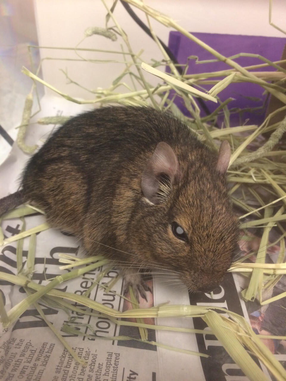
Diabetes mellitus is a chronic endocrine disorder characterised by persistent hyperglycaemia due to impaired insulin production, insulin action or both. This metabolic disorder is particularly predominant in degus (Octodon degus), small rodents native to the regions of Chile (Figure 1). Degus have become popular as pets, due to their social nature and fascinating behaviours. However, their genetic predisposition to diabetes presents unique challenges in veterinary medicine. This article aims to provide a comprehensive overview of diabetes mellitus in degus, covering its aetiology, clinical presentation, diagnostic approaches and management strategies. By enhancing the understanding of this condition, veterinary professionals can improve the care and quality of life for degus affected by this condition.

Aetiology and pathophysiology of diabetes mellitus in degus
Molecular and histological evidence suggests that in these rodents there is a natural resistance to insulin due to their lower metabolic activity and their reduced insulin receptor binding affinity (Ardiles et al., 2013). Degus’ glucose levels are similar to other mammals and the glucose intolerance of the species stems from the different physiological activity of insulin and the general failure of their pancreas (Opazo et al., 2004; Spear et al., 1984a). Type 2 diabetes develops spontaneously in degus due to intolerance to a high-sugar diet and the resulting obesity. This may lead to secondary liver disease, such as hepatic lipidosis (Keeble, 2009).
Degus certainly have a strong predisposition to diabetes as a result of this insulin-reduced activity, but reports on Langerhans islet amyloidosis (Nishi and Steiner, 1990) and cytomegalovirus associated insulitis (Fox and Murphy, 1979) suggest that pancreatic activity in general is compromised in this species.
Genetic predisposition and environmental factors

Degus can develop spontaneous diabetes mellitus, primarily characterised by amyloidosis of the Langerhans islets. Factors contributing to the development of diabetes in degus include cytomegalovirus-induced insulitis, the presence of alpha-cell crystals with herpes-type viruses and diets high in sugar, such as guinea pig chow or fresh fruit, which elevate blood sugar levels (Spear et al., 1984b). Degus are herbivorous rodents anatomically and behaviourally adapted to a diet rich in fibre with moderate-to-low levels of non-structural carbohydrates. In captivity, it is essential that degus are provided with foods that closely mimic the nutritional content of their natural diet. This includes high-quality hay, specific degu pellets and limited amounts of fresh vegetables and fruits to ensure their dietary needs are met (Edwards, 2009).
Clinical signs
Clinical signs of diabetes mellitus in degus are similar to those seen in other mammals, but can be difficult to identify in the early stages: polyuria and polydipsia (Opazo et al., 2004), weight gain due to polyphagia (Edwards, 2009, Bihun and Bauck, 2004), lethargy, cataracts (with impaired vision possibly leading to blindness) (Figure 2) and poor coat condition (unkempt or thin) (Figure 3), as well as dehydration (fluid loss due to urination not being balanced with fluid intake) (Edwards, 2009).

Diagnosis of diabetes mellitus in degus
In the first instance, a thorough history and physical examination are crucial. If clinical signs consistent with diabetes mellitus are found, a fasting blood glucose test can be performed. The mean blood glucose concentration in fasting degus was reported to be 4.34mmol/l (Opazo et al., 2004). Testing urine for glucose and ketones is another useful diagnostic tool for diabetes (Figure 4). The detection of glucose (glycosuria) and ketones (ketonuria) in the urine strongly suggests diabetes in degus. The fructosamine test measures the levels of fructosamine in the blood, which indicates the average blood glucose levels over the past 1 to 2 weeks. Elevated fructosamine levels are indicative of chronic hyperglycaemia and can confirm a diagnosis of diabetes in degus. Finally, the HbA1c (glycated haemoglobin) test measures the percentage of haemoglobin that has glucose attached to it, providing an average blood glucose level over the previous 2 to 3 months. Elevated HbA1c levels indicate poor long-term glucose control, confirming chronic hyperglycaemia (Edwards, 2009).


Treatment
The first, and logical, step of the treatment should be to adapt a balanced diet to the condition of the patient. In order to effectively manage diabetes mellitus in degus, it is crucial to maintain a low-sugar diet by avoiding fruits and commercial treats that are high in sugar. Instead, focus on providing a high-fibre diet that includes hay, fresh vegetables and specially formulated degu pellets. Additionally, it’s important to adhere to a regular feeding schedule, ensuring consistent feeding times to help regulate blood sugar levels.
Medical therapy also plays an essential part in the treatment. First, insulin can be administered at 1 to 3U/kg every 12 to 24 hours SC, with a starting dose of 1U/kg (Carpenter and Harms, 2018). Alternatively, metformin, which works by decreasing glucose production in the liver, improving insulin sensitivity and enhancing peripheral glucose uptake, can be prescribed. No dosage has been reported in degus, but hypoglycaemic effects of metformin are observed at the dose of 250mg/kg orally in mice (Bailey et al., 1986). Consistent monitoring of blood glucose levels is crucial for effectively managing diabetes in degus. Less invasive monitoring can be performed by testing urine samples for glucosuria; however, this is a much less accurate monitoring method.
Prevention of diabetes mellitus in degus
Successfully managing possible cases of diabetes mellitus in degus starts with prevention. Exercise is important, and owners should encourage daily physical activities and create interactive enrichments. Weight management is a necessity, as well as the choice of a balanced and adapted diet. Finally, minimising stressors to prevent the exacerbation of the clinical signs, as well as offering a stable environment to degus, will help in supporting overall health.
Conclusion
In conclusion, diabetes mellitus in degus requires a multifaceted approach to understanding, management and care. Understanding its causes, including genetic and environmental factors, is essential for effective treatment. Early detection through vigilant monitoring and diagnostic testing is crucial. Management focuses on a balanced low-sugar diet, regular exercise, and blood glucose monitoring, with medical treatments as needed. Preventative measures, such as weight management and minimising stress, are key to reducing the risk of diabetes.
By enhancing knowledge and care practices, veterinarians and pet owners can together significantly improve the quality of life for degus with diabetes, ensuring they live healthy and happy lives.
_OL_CourseThumb.jpg?width=500&height=300&name=IVE_GPCert(ExAP)_OL_CourseThumb.jpg)
Exotic Animal Practice
ISVPS General Practitioner Certificate (GPCert)
Postgraduate Certificate (PgC)
 July 2026
 Online
References (click to expand)
| Ardiles, A. O., Ewer, J., Acosta, M. L., Kirkwood, A., Martinez, A. D., Ebensperger, L. A., Bozinovic, F., Lee, T. M. and Palacios, A. G. | 2013 | Octodon degus (Molina 1782): a model in comparative biology and biomedicine. Cold Spring Harbour Protocols, 4 |
| Bailey, C. J., Flatt, P. R. and Ewan, C. | 1986 | Anorectic effect of metformin in lean and genetically obese hyperglycaemic (ob/ob) mice. Archives Internationales de Pharmacodynamie et de Thérapie, 282, 233-239 |
| Bihun, C. and Bauck, L. | 2004 | Small rodents: basic anatomy, physiology and clinical techniques. In: Quesenberry, K. E. and Carpenter, J. W. (eds), Ferrets, Rabbits and Rodents: Clinical Medicine and Surgery, 2nd ed., Elsevier, pp.286 |
| Carpenter, J. W. and Harms, C. | 2018 | Carpenter’s Exotic Animal Formulary, 6th ed. Elsevier Health Sciences |
| Edwards, M. S. | 2009 | Nutrition and behavior of degus (Octodon degus). Veterinary Clinics of North America: Exotic Animal Practice, 12, 237-253 |
| Fox, J. G. and Murphy, J. C. | 1979 | Cytomegalic virus-associated insulitis in diabetic Octodon degus. Veterinary Pathology, 16, 625-628 |
| Keeble, E. | 2009 | Rodents: biology and husbandry. In: Keeble, E. and Meredith, A. (eds), BSAVA Manual of Rodents and Ferrets, 1st ed. BSAVA, Gloucester |
| Nishi, M. and Steiner, D. F. | 1990 | Cloning of complementary DNAs encoding islet amyloid polypeptide, insulin, and glucagon precursors from a new world rodent, the degu, Octodon degus. Molecular Endocrinology, 4, 1192-1198 |
| Opazo, J. C., Soto-Gamboa, M. and Bozinovic, F. | 2004 | Blood glucose concentration in free-ranging degus, Octodon degus (Rodentia: Octodontidae): is there evidence of a homeostatic set point? Comparative Biochemistry and Physiology Part A: Molecular and Integrative Physiology, 137, 251-257 |
| Spear, G. S, Caple, M. V. and Sutherland, L. R. | 1984a | The pancreas in the degu. Experimental and Molecular Pathology, 40, 295-310 |
| Spear, P. A., Austad, S. N. and Kenagy, G. J. | 1984b | Comparative glucose tolerance of two hystricomorph rodents, Octodon degus and Cavia porcellus. Journal of Comparative Physiology B, 154, 477-481 |