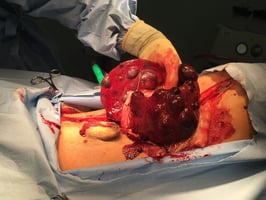
Emergency splenectomy is a common and often life-saving procedure in small animal practice, most frequently performed in dogs presenting with haemoperitoneum. Rapid recognition and surgical intervention are critical, particularly in cases of ongoing intra-abdominal haemorrhage.
This video, recorded by Jon Hall FRCVS, demonstrates how to perform an emergency splenectomy on a cadaver.
When to perform an emergency splenectomy
An emergency splenectomy should be considered in dogs with suspected or confirmed splenic disease causing haemodynamic instability, particularly where there is evidence of active intra-abdominal bleeding.
Common indications include:
- Haemoperitoneum of suspected splenic origin, especially in unstable patients where rapid surgical intervention is required
- Ruptured splenic mass, whether malignant (e.g. haemangiosarcoma) or benign (e.g. haematoma)
- Splenic torsion, which may present with acute abdominal distension, pain, and cardiovascular compromise
- Severe abdominal trauma involving the spleen
- Ongoing blood loss despite stabilisation, where medical management alone is insufficient
In many cases, a definitive diagnosis is not available prior to surgery, and splenectomy is then both a diagnostic and therapeutic procedure, aimed at controlling haemorrhage and stabilising the patient.
Step-by-step guide to emergency splenectomy surgery
Step one: Patient prep
The patient will need to be placed under a full general anaesthetic - they will therefore need to be stabilised (where possible), and may require:
-
Blood transfusions
-
Multimodal pain relief
- Intravenous fluids
The abdomen should have a wide clip and be aseptically prepared.
Step two: Opening the abdomen
The initial incision should be on the ventral midline, from the xiphoid process to the pubis. Once through the skin and subcutis, a self-retaining retractor can help to allow proper exposure. The spleen is found in the cranial left quadrant, near the fundus of the stomach.
Tips for splenectomy
1. When ligating the splenic artery and vein (central vessels), do not occlude the blood supply to the pancreas
2. Double ligate all major vessels
3. Carefully inspect all ligations for evidence of haemorrhage before and during closure
Step three: Closing the abdomen
Before closure, all swabs should be removed and counted to make sure none are left behind. The abdomen should be closed in a routine fashion.
Author
Jon Hall, MA, VetMB, CertSAS, DipECVS, SFHEA, FRCVS, is a European and RCVS Recognised Specialist in Small Animal Surgery. Jon is head of Soft Tissue Surgery at Wear Referrals and a Professor of Small Animal Surgery at the University of Nottingham.