Case study: acute collapse and haemorrhagic diarrhoea in a Poodle

It's 7am on a Saturday morning and a collapsed Standard Poodle is on their way down... can you solve the case and save the dog?

feature-img

It's early on a Saturday morning, and your first call has come in. A 5-year-old Standard Poodle with vomiting and diarrhoea is on its way, collapsed.

Think you can solve the case? Grab a pen and paper, set a timer (to record your CPD!) and settle down...

Presentation

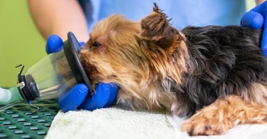
A 5-year-old neutered male Standard Poodle is presented as an emergency appointment by the owner of a local boarding kennels. He has been boarding there for the last four days while his owners are away.

The kennel owner reports that the dog was quiet yesterday evening and refused his dinner. Overnight, he passed several episodes of diarrhoea, which became haemorrhagic by the morning. He has vomited twice since breakfast and is now weak, dull and reluctant to stand.

The kennel owner is concerned about “something infectious”. There are other dogs on site, and although no other dogs are currently unwell there was one with mild diarrhoea last week. The patient is reported to be fully vaccinated according to the kennels’ admission record, but the kennel owner does not have access to his full medical history.

On arrival, the dog is quiet, weak and ambulatory only with support. He is taken straight through for triage while you contact the owner.

What additional history would you like from the kennel owner and dog owner?
Note down your questions, then click 'see more' for the answers.

The kennel owner confirms that the dog had been eating normally until the previous evening. There has been no known dietary indiscretion while in kennels. He has not received any medication while boarding. There has been no known toxin exposure. His diarrhoea has been frequent, watery and blood-stained, with some fresh blood and mucus noted in the most recent episode.

The dog has not been observed urinating that morning, but the kennel owner is unsure whether this is abnormal. Water intake has not been closely monitored.

Owner history

The owner is contacted by telephone. They report that the dog is greedy and "sensitive-stomached", so he's prone to diarrhoea when he gets food he shouldn't. This usually resolves spontaneously or after symptomatic treatment if he's been lethargic. When asked if he would eat something he shouldn't, they answered "most definitely - we don't let him have toys because he'd eat them" and he would "eat all sorts of disgusting things if given half a chance". They don't know if he had anything he shouldn't, but admit they wouldn't have noticed if something small - like a pair of socks - went missing in the hurry to pack.

The owner does not think he has been drinking or urinating excessively over the last few weeks. There is no known history of seizures, collapse, or toxin exposure. He is not receiving any long-term medication other than his flea and worm tablets, which he had last week. Vaccination is up to date.

They are a bit annoyed at the intrusion on their holiday, feeling that the dog is often a bit quiet while in kennels, so this is a regular issue that the kennel owner should have been able to deal with without needing to see the vet.

See more See less


Clinical examination

Explaining to the owner that from triage the dog does in fact need to see a vet this morning, you continue your examination.

  • On initial examination, the dog is dull and weak but responsive. He is able to lift his head and interact briefly, but quickly becomes recumbent again.

  • His mucous membranes are bright red and tacky, with a capillary refill time of approximately 2.5 seconds. Peripheral pulses are weak and slightly thready. His extremities feel cool.

  • Heart rate is 92 beats per minute with a regular rhythm. No murmur is detected. 

  • Respiratory rate is 28 breaths per minute, with no obvious increased respiratory effort. Lung sounds are unremarkable.

  • Rectal temperature is 37.7°C.

  • Abdominal palpation reveals mild, diffuse discomfort but no obvious focal pain, mass effect or abdominal distension. The bladder cannot be felt. There is faecal staining around the perineum, consistent with the reported haemorrhagic diarrhoea.

  • A skin tent is present

  • Body condition score is 4/9 and weight is 28.4kg.

What is your problem list for this dog?

The initial problem list includes:

  • Acute weakness/collapse
  • Haemorrhagic diarrhoea
  • Vomiting
  • Dehydration
  • Mild abdominal discomfort
  • Inappropriately low heart rate 
  • Low body temperature
  • Known foreign body risk

Note: The heart rate should not be interpreted in isolation. A heart rate of around 90 beats per minute may be within a reference interval for a dog of this size, but it is inappropriate for a collapsed, poorly perfused patient. 

See more See less


For each problem, write a differential diagnosis list.

Circle the ones that cover multiple problems as priorities, but don't forget that more than one problem could explain the signs.

Diagnoses shown in bold are those that appear in several of the problem categories below and are therefore high-priority, but you should remember that more than one problem could be causing the signs.

Acute weakness/collapse

Differential diagnoses include:

  • Hypoadrenocorticism/Addisonian crisis
  • Acute haemorrhagic diarrhoea syndrome
  • Sepsis or systemic inflammatory response syndrome
  • Acute kidney injury or uraemic crisis
  • Hypoglycaemia
  • Electrolyte disturbance, particularly hyperkalaemia or hyponatraemia
  • Cardiac arrhythmia
  • Toxin exposure

Haemorrhagic diarrhoea

Differential diagnoses include:

  • Acute haemorrhagic diarrhoea syndrome
  • Infectious enteritis
  • Dietary indiscretion
  • Gastrointestinal foreign body or obstruction
  • Hypoadrenocorticism/Addisonian crisis
  • Pancreatitis
  • Gastrointestinal ulceration
  • Coagulopathy

Vomiting

Differential diagnoses include:

  • Acute haemorrhagic diarrhoea syndrome
  • Dietary indiscretion
  • Gastrointestinal foreign body or obstruction
  • Pancreatitis
  • Hypoadrenocorticism/Addisonian crisis
  • Acute kidney injury or uraemia
  • Toxin exposure
  • Infectious enteritis

Dehydration

Differential diagnoses include:

  • Gastrointestinal fluid loss secondary to vomiting and diarrhoea
  • Acute haemorrhagic diarrhoea syndrome
  • Hypoadrenocorticism/Addisonian crisis
  • Acute kidney injury
  • Sepsis or systemic inflammatory response syndrome
  • Reduced water intake due to illness or kennel stress
  • Diabetes mellitus or another cause of osmotic diuresis
  • Pancreatitis

Mild abdominal discomfort

Differential diagnoses include:

  • Acute haemorrhagic diarrhoea syndrome
  • Infectious enteritis
  • Pancreatitis
  • Gastrointestinal foreign body or obstruction
  • Hypoadrenocorticism/Addisonian crisis
  • Uroabdomen or urinary tract disease
  • Peritonitis
  • Hepatobiliary disease

Low heart rate

Differential diagnoses include:

  • Hypoadrenocorticism/Addisonian crisis
  • Hyperkalaemia
  • Cardiac arrhythmia or conduction disturbance
  • Toxin exposure
  • Increased vagal tone secondary to gastrointestinal disease
  • Severe systemic illness (eg sepsis) with impending cardiovascular decompensation
  • Drug effect, although there is no known medication history at this stage
  • Primary cardiac disease


Well done if your differential list had some or most of the above. It's important to keep an open mind at this point!

See more See less


Next steps

What are your immediate priorities for this patient?

This dog is clinically unstable. The immediate priority is not to reach a definitive diagnosis, but to support perfusion, identify immediately life-threatening abnormalities, and collect key samples before treatment alters the results.

The dog is moved to the emergency treatment area. 

Initial stabilisation should include:

  • Rapid assessment of airway, breathing and circulation
  • Oxygen supplementation if there is any concern about oxygen delivery, respiratory effort or worsening shock
  • Intravenous access, ideally with a large-bore catheter
  • Collection of blood for point-of-care testing and possible endocrine testing before steroid administration
  • Active warming, as the dog is low-normal in temperature and peripherally cool
  • Fluid resuscitation
  • Isolation or barrier nursing precautions while infectious gastrointestinal disease remains possible

This patient should be reassessed frequently. Useful reassessment points include:

  • Mentation
  • Heart rate and rhythm
  • Pulse quality
  • Mucous membrane colour
  • Capillary refill time
  • Extremity temperature
  • Respiratory rate and effort
  • Blood pressure
  • Repeat lactate if initially increased
  • Urine production once resuscitation is underway

Analgesia and antiemetics should also be considered as part of the initial stabilisation, depending on the patient and your priorities.

At this stage, empirical antibiotics are not automatic. They may be indicated if there is evidence of sepsis, neutropenia, septic peritonitis, bacterial translocation with systemic compromise, or another clear infectious focus. However, haemorrhagic diarrhoea alone is not a reason to give antibiotics without considering the wider clinical picture.

If you didn't get this right, or felt not confident, make a note of the gaps in your knowledge so you can plan more CPD in this area

See more See less

Fluid considerations

What fluids do you want to give this patient? What rate would be suitable?

This dog needs fluids for resuscitation.

He is weak, poorly perfused, hypotensive, dehydrated and hyperlactataemic, so an isotonic crystalloid bolus is appropriate while further assessment and point-of-care testing are underway.

A practical initial plan would be:

  • Place IV access, ideally with a large-bore catheter.
  • Start an isotonic crystalloid bolus.
  • Give 10–20 ml/kg IV over 10–15 minutes. In this dog, that equates to 284-568ml of fluids.
  • Reassess after each bolus rather than prescribing a fixed “shock dose”.
  • Repeat boluses if perfusion remains poor, while monitoring closely for response and fluid overload.

Suitable fluid choices include:

  • A balanced isotonic crystalloid, such as Hartmann’s solution/compound sodium lactate or Plasma-Lyte

Do not add potassium to the fluids. The priority at this stage is timely restoration of circulating volume, with frequent reassessment.

Reassessment should include:

  • Mentation
  • Heart rate and rhythm
  • Pulse quality
  • Bodyweight
  • Mucous membrane colour
  • Capillary refill time
  • Extremity temperature
  • Respiratory rate and effort
  • Blood pressure
  • Lactate
  • Urine production once resuscitation is underway
  • Repeat electrolytes and glucose once available

If the dog improves after a bolus, fluid therapy can then be adjusted to account for ongoing deficits, maintenance requirements and continuing gastrointestinal, urinary, and respiratory losses.

If the dog fails to respond as expected, or deteriorates despite repeated boluses, the plan should be reassessed.

See more See less


What tests do you want to run on this dog?

Think about which are priorities and which could be safely delayed

Immediate point-of-care tests

The first tests should aim to identify life-threatening abnormalities and guide resuscitation. These include:

  • PCV and total solids
  • Blood glucose
  • Electrolytes, especially sodium, potassium and chloride
  • Venous blood gas and acid-base status, if available
  • Lactate
  • Blood pressure
  • ECG
  • Packed cell volume/total solids reassessment if ongoing haemorrhagic diarrhoea is significant
  • POCUS

These results help assess perfusion, hydration, electrolyte derangements, acid-base status, renal involvement and the risk of clinically significant arrhythmia.

Minimum database

As soon as practical, the dog should have:

  • CBC and smear analysis
  • Serum biochemistry
  • Basal cortisol
  • Urinalysis, including urine specific gravity
See more See less

Test results

When you've decided which tests you want to run, click below to see the results

The dog remains dull but responsive while initial fluid resuscitation is underway. After a 10 ml/kg isotonic crystalloid bolus, his pulse quality improves slightly, but his mucous membranes remain tacky and his extremities remain cool. 

ECG

ECG_CaseStudy

 The rhythm is regular and sinus in origin, with a heart rate of approximately 90 beats per minute. P waves are present and the QRS complexes are narrow. The T waves are subjectively tall and slightly tented, but there is no widening of the QRS complexes, loss of P waves or obvious bradyarrhythmia at this stage. 

Lactate

Lactate is 5.6mmol/L (Ref: <2.5mmlol/L)

PCV and Total Solids

PCV: 53% (37-55%)

Total solids: 59g/L (55-75g/L)

Blood pressure

Doppler systolic blood pressure after the first fluid bolus is 88mmHg.

The dog remains hypotensive after the initial fluid bolus. This supports ongoing cardiovascular compromise and indicates that further reassessment-guided resuscitation is needed. 

Haematology

Parameter Value Reference
RBC 8.1 5.39–8.70
Haematocrit 52.0 38.3–56.5
Haemoglobin 18.4 13.4–20.7
MCV 64.6 59–76
MCH 22.9 21.9–26.1
MCHC 35.4 32.6–39.2
% Reticulocyte 0.6
Reticulocytes 48.0 10–110
Reticulocyte haemoglobin 25.8 24.5–31.8
WBC 8.4 4.9–17.6
% Neutrophils 64.3
% Lymphocytes 25.0
% Monocytes 6.0
% Eosinophils 4.8
% Basophils 0.0


Manual blood smear review is performed.

  • Platelet estimate: adequate, with occasional small platelet clumps.
  • Red blood cell morphology: unremarkable. No spherocytes, schistocytes, Heinz bodies or haemoparasites seen.
  • White blood cell morphology: predominantly mature neutrophils.
  • Left shift: not significant.
  • Toxic change: not seen.
  • Lymphocytes and eosinophils are present in numbers that are not typical of a marked stress leukogram.
  • No organisms seen.

Biochemistry

Parameter Value Ref  
Glucose 3.6 3.5–6.5 Normal
SDMA 18 0–14 High
Creatinine 186 40–130 High
Urea 16.8 3.0–9.0 High
Phosphate 1.9 0.8–1.8 High
Calcium 2.88 2.2–2.8 High
Total protein 61 55–75 Normal
Albumin 31 26–38 Normal
Globulin 30 22–40 Normal
Albumin:Globulin ratio 1.0 0.7–1.5 Normal
ALT 48 10–90 Normal
AST 42 16–55 Normal
ALP 34 15–150 Normal-low
GGT 2 0–13 Normal
Bilirubin 3 0–10 Normal
Cholesterol 3.1 3.2–7.0 Low
Basal cortisol <0.2 2.0-6.0 Low


Electrolytes and venous blood gas 

Parameter Value Ref  
Sodium 130 140–155 Low
Potassium 5.9 3.5–5.5 High
Na:K ratio 22 >27 Low
Chloride 96 105–120 Low
pH (venous) 7.21 7.35–7.45 Low
pCO2 36 35-45 Normal
Bicarbonate 14.6 18–24 Low
Base excess -12 -4 to +4 Low
Anion gap 21 12–24 High-normal


POCUS

No significant free abdominal fluid is identified. A very small volume of anechoic fluid is seen between intestinal loops, but this is considered non-specific and insufficient for sampling. 

Urinalysis

No bladder can be felt on palpation and there is little urine on ultrasound. The decision is made to not attempt urinalysis until later.

See results See less

Differential diagnoses

Look again at your differential diagnosis list... is there anything you can rule out or increase suspicion for at this point?

The minimum database significantly changes the weighting of the differential diagnosis list.

The combination of an inappropriately normal heart rate, hypotension, hyponatraemia, hyperkalaemia, hypochloraemia, azotaemia, low-normal glucose, mild hypercalcaemia, low cholesterol, low basal cortisol and absence of a convincing stress leukogram makes hypoadrenocorticism/Addisonian crisis the leading differential.

However, the dog also has acute haemorrhagic diarrhoea, vomiting, mild abdominal discomfort and azotaemia. Several emergency differentials remain plausible and clinically important.

A reasonable prioritised list at this stage would be:

1. Hypoadrenocorticism/Addisonian crisis

This is now the leading differential.

Supportive features include:

  • Recurrent vague gastrointestinal signs
  • Lower body condition score
  • Collapse and poor perfusion
  • Inappropriately normal heart rate for degree of shock
  • Hyponatraemia
  • Hyperkalaemia
  • Hypochloraemia
  • Azotaemia
  • Hypoglycaemic for state of stress
  • Mild hypercalcaemia
  • Low cholesterol
  • Low basal cortisol
  • Lack of a clear stress leukogram despite significant illness

The haemorrhagic diarrhoea does not rule this out. Dogs with hypoadrenocorticism may present with acute gastrointestinal signs, including severe diarrhoea, and can be mistaken for primary gastrointestinal disease.

2. Acute haemorrhagic diarrhoea syndrome with hypovolaemic shock

This remains plausible because the dog has acute haemorrhagic diarrhoea, vomiting, dehydration and shock.

However, it does not fully explain the recurrent history, the inappropriately normal heart rate, the electrolyte pattern or the low basal cortisol. It may still be present as a concurrent or secondary process, but it is now less satisfying as the sole diagnosis.

3. Acute kidney injury or mixed pre-renal/renal azotaemia

Azotaemia may be pre-renal due to hypovolaemia and poor perfusion. However, acute kidney injury remains possible, particularly in a collapsed dog with vomiting, diarrhoea and possible hypotension.

The hyperkalaemia could fit renal compromise, but the electrolyte pattern and low cortisol increase suspicion that the azotaemia may be part of an Addisonian crisis rather than primary renal disease.

Urinalysis will be important.

4. Gastrointestinal foreign body or obstruction

A gastrointestinal foreign body or obstruction remains possible because of vomiting, abdominal discomfort and systemic compromise.

The haemorrhagic diarrhoea is less typical for a simple obstruction but does not exclude it. Imaging is needed before this can be safely deprioritised, especially given the incomplete kennel history.

5. Pancreatitis

Pancreatitis could explain vomiting, abdominal discomfort, lethargy, dehydration and shock. It can also occur alongside other disease processes.

However, the electrolyte abnormalities, low cortisol and lack of a stress leukogram make pancreatitis less likely as the primary explanation. It remains a differential to investigate, particularly if imaging or pancreatic testing later supports it.

6. Sepsis or systemic inflammatory response syndrome

Sepsis remains a critical differential in any shocked patient with gastrointestinal signs. The kennel environment and haemorrhagic diarrhoea may also raise concern for infectious disease.

At present, there is no pyrexia, neutropenia, marked inflammatory leukogram, toxic change or clear septic focus. Sepsis is therefore not the leading diagnosis, but it cannot be dismissed yet.

7. Uroabdomen or urinary tract disease

Hyperkalaemia and azotaemia mean urinary tract disease should still be considered, even though the bladder was small and soft on palpation.

Uroabdomen is less likely without abdominal distension or free fluid suspicion, but imaging and urinalysis are needed to reduce this concern.

8. Toxin exposure

Toxin exposure remains possible because the history from the kennel is incomplete. Some toxins could cause vomiting, diarrhoea, collapse, electrolyte disturbance or renal injury.

There is no specific exposure history at this stage, so this is lower on the list, but it should remain in the background until the case becomes clearer.

See more See less


Further testing

Following two fluid boluses of 350ml, the dog is brighter. He can now maintain sternal recumbency without support and briefly wags his tail when handled. His mucous membranes are pinker and less tacky, with a CRT of 2s and his peripheral pulse quality has improved. 

During this period, he passes another small volume of watery haemorrhagic diarrhoea. No urine has yet been obtained.

The response to fluids is encouraging, but incomplete. His lactate has improved but remains increased, his blood pressure remains marginal, and his heart rate remains inappropriately normal for a dog that is still not fully perfused.

Adjust your differential list if needed, then move on to further testing...

What are your next steps?

Further testing should be targeted. The aim is not to “do everything”, but to answer the questions that remain clinically important after the minimum database.

At this point, the most useful next steps are:

Urinalysis

Urinalysis is a priority because the dog is azotaemic and mildly hyperkalaemic.

The key questions are:

  • Is the urine appropriately concentrated for the degree of azotaemia?
  • Is there evidence of urinary tract inflammation, infection, haematuria or pigmenturia?
  • Are there ketones or glucosuria?

A urine specific gravity that is not appropriately concentrated in an azotaemic patient would prevent a simple “dehydration only” interpretation, although results must be interpreted in light of fluid therapy already given.

Abdominal imaging

Further abdominal ultrasound because several important differentials remain possible.

The key questions are:

  • Are there changes suggestive of pancreatitis?
  • Is there severe intestinal wall thickening, loss of layering or concern for peritonitis?
  • Is there evidence of abdominal neoplasia or another structural disease?
  • Is there evidence of a foreign body?
  • Can the adrenal glands be visualised, and are they subjectively small?

However, imaging should be brief and focused if the patient remains unstable.

See more See less

Test results

Further ultrasound scan

The urinary bladder is small to moderately filled and has a normal wall thickness. No obvious bladder wall defect is seen. A urine sample is collected by cystocentesis.

Both kidneys are visualised. They are normal in size and shape, with no renal pelvic dilation. There is no evidence of ureteral obstruction. A quick look at the intestines shows no sign of foreign body, no plication, and no obstructive pattern. The pancreas appears normal in size and homogenicity, but the dog begins to appear stressed and you make the decision not to scan any further. 

Urinalysis

The urine dipstick shows a trace of protein and a trace of blood, and is otherwise negative. The SG is 1.021.

Sediment: inactive; RBC 0–3/hpf, WBC 0–2/hpf, no bacteria seen, no casts. 


See more See less


Narrowing your differentials...

With those further tests in mind, your differential diagnosis list should be shorter. 

What is your new differential diagnosis list?

Based on the history, clinical examination, minimum database, response to initial stabilisation, POCUS and urinalysis, the current differential diagnosis list is:

  • Hypoadrenocorticism/Addisonian crisis
    This is now the leading differential. It best explains the recurrent vague gastrointestinal history, recent weight loss, collapse, inappropriately normal heart rate, hyponatraemia, hyperkalaemia, hypochloraemia, azotaemia, low-normal glucose, mild hypercalcaemia, low cholesterol, low basal cortisol and absence of a convincing stress leukogram.
  • Acute haemorrhagic diarrhoea syndrome or severe enteritis
    The dog has acute haemorrhagic diarrhoea, vomiting, dehydration and hypovolaemia. However, it does not fully explain the weight loss, electrolyte pattern, low basal cortisol or inappropriately normal heart rate.
  • Acute kidney injury or mixed pre-renal/renal azotaemia
    Azotaemia remains clinically relevant. The urine specific gravity is not appropriately concentrated for the degree of azotaemia, although interpretation is complicated by prior fluid boluses and the effects of hypoadrenocorticism on renal concentrating ability.
  • Pancreatitis
    Pancreatitis could still contribute to vomiting, abdominal discomfort and systemic compromise.

Lower-priority differentials

  • Sepsis or systemic inflammatory response syndrome
    Sepsis remains a consideration in any shocked patient with gastrointestinal signs. However, there is no pyrexia, neutropenia, toxic change, marked inflammatory leukogram, septic focus or convincing evidence of peritonitis at this stage.
  • Gastrointestinal obstruction or foreign body
    This is less likely because POCUS did not show an obstructive pattern, marked segmental intestinal dilation, plication or visible foreign material. However, you shouldn't rule it out completely without additional testing, as your scan was incomplete
  • Uroabdomen or urinary tract obstruction
    This is now unlikely. The bladder and proximal urethra are not suggestive of obstruction, there is no significant abdominal effusion, and the kidneys do not show obstructive change.
  • Primary urinary tract infection
    This is unlikely based on the inactive urine sediment, absence of bacteria seen in-house, and lack of supportive urinary tract findings.
  • Toxin exposure
    This remains a lower-priority differential because the kennel history is incomplete. There is currently no specific exposure history or diagnostic finding that strongly supports it.

Overall, the case now most strongly supports hypoadrenocorticism/Addisonian crisis, although ongoing monitoring and confirmatory endocrine testing are still needed before the diagnosis can be considered definitive.

See more See less


What do you want to do next?

Given your differential diagnosis list, an ACTH stimulation test is a sensible move. 

A pre-ACTH blood sample is collected, and synthetic ACTH is administered intravenously. The post-ACTH sample is planned for 60 minutes later.

Fifteen minutes later, the nurse alerts you that the dog has suddenly become more obtunded and that his heart rate has dropped on the monitor.

On reassessment, he is no longer able to maintain sternal recumbency. His mucous membranes are pale pink, his extremities are cold, and his femoral pulses are weak.

Repeat parameters show:

Parameter Result
Heart rate 54 bpm
Respiratory rate 24 breaths/min
Doppler systolic blood pressure 66 mmHg
Temperature 37.5°C
Blood glucose 2.9 mmol/L
Lactate 6.5 mmol/L
Venous pH 7.16
Bicarbonate 12.8mmol/L
Base excess -16 mmol/L


You obtain a repeat ECG. It shows a bradyarrhythmia with tall, tented T waves. P waves are difficult to identify consistently, and the QRS complexes appear slightly wider than on the initial ECG.

Given the ECG changes, you immediately recheck electrolytes.

Parameter Previous result Repeat result
Sodium 130 mmol/L 128 mmol/L
Potassium 5.9 mmol/L 7.3 mmol/L
Chloride 96 mmol/L 94 mmol/L


The dog is now hypotensive, hypoglycaemic, significantly hyperkalaemic and more acidotic, with ECG changes consistent with clinically important hyperkalaemia.

See more See less


What should be your immediate priorities?

This dog is now unstable and has life-threatening hyperkalaemia with ECG changes. The immediate priority is emergency treatment, not completion of the ACTH stimulation test. The team should avoid assuming this deterioration is a reaction to ACTH or handling. 

The post-ACTH cortisol sample should still be collected at the correct time if this can be done without interfering with resuscitation, but you should not wait for that sample before treating the dog.

A practical immediate plan would be:

  • Continue ECG monitoring
  • Administer calcium gluconate for cardioprotection
  • Give dextrose and consider insulin/dextrose therapy to shift potassium intracellularly
  • Continue reassessment-guided isotonic crystalloid resuscitation
  • Administer dexamethasone
  • Recheck potassium, glucose, lactate and blood pressure after treatment
  • Collect the post-ACTH cortisol sample at 60 minutes if doing so does not compromise emergency care

Results

Over the next 45–60 minutes, the dog begins to improve. He becomes brighter and is again able to maintain sternal recumbency. His pulse quality improves, his mucous membranes become pinker, and his extremities feel warmer. 

Repeat ECG shows a regular sinus rhythm. P waves are now visible, the QRS complexes are narrow, and the T waves are less prominent than during the deterioration. 

You are able to collect the second ACTH stimulation test sample:

Post-ACTH cortisol remains low, confirming no appropriate adrenal response. The dog has Addison's disease (hypoadrenocorticism).

See more See less


The next 48h...

You may have a diagnosis, but the dog isn't out of the woods yet!

What are the key points for treating and monitoring this dog over the next 24-48 hours?

This dog should remain hospitalised and closely monitored.

Supportive care should continue as needed, including fluids, antiemetic treatment, gastroprotectant treatment if clinically justified, warmth, nursing care and nutritional support once vomiting is controlled.

Glucocorticoid replacement should also continue - usually dexamethasone to begin with, transitioning to oral prednisolone when the dog is eating.

Mineralocorticoid replacement should now be started, unless there is a specific reason to delay.

See more See less


The owner is on the phone demanding an update...

What do you tell the owner about their dog's diagnosis and the plan?

Think about the diagnosis, current status, prognosis as well as your treatment plan. 

  • Diagnosis
    • The ACTH stimulation test confirms hypoadrenocorticism, also known as Addison’s disease.
    • He has presented in an Addisonian crisis, which is a life-threatening emergency.
  • Current status
    • He has improved after emergency treatment.
    • His heart rhythm, blood pressure, potassium and blood glucose are better than they were
    • He is still not stable enough to go home.
  • Prognosis over the next 24-48 hours
    • Guarded to fair at this stage, improving if he continues to respond to treatment.

  • Treatment over the next 24–48 hours
    • Continue intravenous fluids to support blood pressure, hydration, kidney perfusion and electrolyte correction.
    • Continue dexamethasone initially for glucocorticoid replacement and crisis support.
    • Start mineralocorticoid treatment to control sodium, potassium and fluid balance.
    • Continue supportive care for vomiting, diarrhoea, temperature and nutrition as needed.
  • Long-term prognosis
    • Good once stabilised and appropriately treated.
    • He will need ongoing medication and regular blood tests.
    • Medication doses may need adjustment, especially in the first few weeks to months.
See more See less


48 hours later...

Great news - your patient is far better and ready for discharge. You handed to the day team on Monday morning, and they have made an ongoing plan for treatment and discharge.

Well done, you’ve worked through one Addisonian crisis, but this case also used skills from several emergency domains: shock, ECG interpretation, electrolytes, POCUS, endocrine emergencies and GI emergencies. If you found yourself hesitating at any point, that is exactly what structured emergency medicine CPD is designed to fix. 

Now, don't forget to log your CPD and reflect on what you learned today! If this case showed holes in your knowledge, make a plan to fix them. 

References (click to expand)

Resources

References

  • Bugbee A, Rucinsky R, Cazabon S, et al. 2023 AAHA selected endocrinopathies of dogs and cats guidelines. Journal of the American Animal Hospital Association. 2023;59(3):113–135. doi:10.5326/JAAHA-MS-7368
    Useful for current consensus guidance on recognition, diagnosis, treatment and monitoring of canine hypoadrenocorticism.
  • Guzmán Ramos PJ, Bennaim M, Shiel RE, Mooney CT. Diagnosis of canine spontaneous hypoadrenocorticism. Canine Medicine and Genetics. 2022;9:6. doi:10.1186/s40575-022-00119-4
    Useful for diagnostic reasoning, clinical presentation, classification and clinicopathological features.
  • Gold AJ, Langlois DK, Refsal KR. Evaluation of basal serum or plasma cortisol concentrations for the diagnosis of hypoadrenocorticism in dogs. Journal of Veterinary Internal Medicine. 2016;30(6):1798–1805. doi:10.1111/jvim.14589
    Useful for basal cortisol interpretation and why a low basal cortisol is not, by itself, definitive.
  • Lennon EM, Boyle TE, Hutchins RG, Friedenthal A, Correa MT, Bissett SA. Use of basal serum or plasma cortisol concentrations to rule out a diagnosis of hypoadrenocorticism in dogs: 123 cases (2000–2005). Journal of the American Veterinary Medical Association. 2007;231(3):413–416.
    Useful for the principle that basal cortisol is most valuable as a rule-out screening test.
  • Lathan P, Thompson AL. Management of hypoadrenocorticism (Addison’s disease) in dogs. Veterinary Medicine: Research and Reports. 2018;9:1–10. doi:10.2147/VMRR.S125617
    Useful for acute crisis management, long-term glucocorticoid/mineralocorticoid treatment and owner follow-up.
  • Kintzer PP, Peterson ME. Treatment and long-term follow-up of 205 dogs with hypoadrenocorticism. Journal of Veterinary Internal Medicine. 1997;11(2):43–49.
    Useful for long-term prognosis and outcome discussion.
  • Tag TL, Day TK. Electrocardiographic assessment of hyperkalemia in dogs and cats. Journal of Veterinary Emergency and Critical Care. 2008;18(1):61–67. doi:10.1111/j.1476-4431.2007.00268.x
    Useful for the ECG/hyperkalaemia component and the reminder that ECG findings do not always map neatly to serum potassium concentration.
  • Iimori Y, Johnson PA, Thomovsky EJ, Brooks AC, Aghili A. Emergency management of hyperkalemia in dogs and cats — Part 1: Pathophysiology and etiology. Canadian Veterinary Journal. 2026;67(4):448–457.
    Useful for causes of hyperkalaemia, including hypoadrenocorticism, AKI and urinary tract obstruction/rupture.
  • Iimori Y, Johnson PA, Thomovsky EJ, Brooks AC, Aghili A. Emergency management of hyperkalemia in dogs and cats — Part 2: Diagnosis and treatment. Canadian Veterinary Journal. 2026;67(5):563–571.
    Useful for ECG-guided stabilisation, calcium gluconate, insulin/dextrose, glucose monitoring and cause-directed treatment.

Further reading

IVE_GPCert(EM&S)_OL_CourseThumb
online

Emergency Medicine and Surgery

ISVPS General Practitioner Certificate (GPCert)

Postgraduate Certificate (PgC)

date Nov 2026 location Online