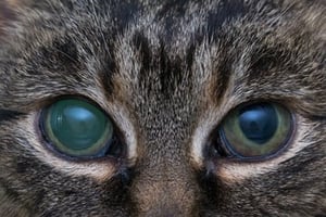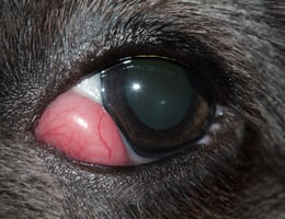
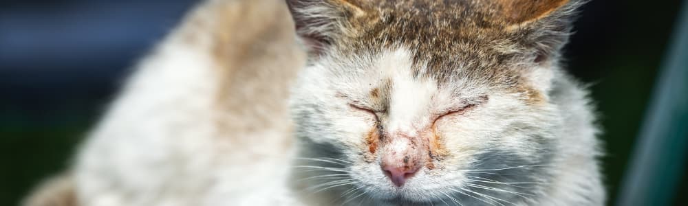
Case study: The cat with 'sore eyes'

Next up on your Wednesday morning list is a 9-year-old cat with 'sore eyes again'. Can you solve this interactive ophthalmology case study?

feature-img

It is a Wednesday morning, and you are halfway through your appointments. Next up is a 9-year-old cat with "sore eyes again".

Think you can help? Grab a pen and paper, set a timer (so you can record your CE!) and settle down with our clinical case. We expect it to take an hour or two to work through properly, but you can always pause and return if you need to.

Presenting problem and initial history

You quickly review the recent history. The cat is a neutered male domestic shorthair. He lives with three other cats, and was adopted after the owner found him as a stray about 5 years ago. He is estimated to be around 9 years old. His last appointment was his vaccination about eight months ago, when a dental procedure was recommended. Before that, he has a general history of cat bite abscesses and recurring upper respiratory tract symptoms.

The owner reports that his "URI is back." Both eyes have been "weepy and irritated" for around five days. The discharge was initially clear, but has become slightly thicker over the last 24-48 hours. The cat has been squinting intermittently, especially in bright light, and has been rubbing his face on the sofa more than usual. He is snuffly too, with some discharge around his nose.

The owner is not especially worried. He has had occasional mild flare-ups of sneezing and watery eyes in the past, usually settling without treatment or with oxytetracycline/polymyxin B ophthalmic ointment. The owner suspects this is another episode. She is a bit annoyed that the receptionist would not give her any more medicine without the cat being seen.

What further information would you like from the owner? 

Have a think, then click below to see the answers

General demeanor, appetite, thirst, and litter box habits: He has been a little quieter than usual at home and has been off his food a little bit, although he is still eating and prefers softer food. There may have been a small amount of weight loss, but he is also "getting older". There is no change in his drinking noted, but she does not know about toileting as he spends a lot of time outdoors. She has noted no vomiting or diarrhea.

History of the problem: Both eyes seemed to become red at roughly the same time. The owner does not think one eye was affected first, although the right eye may look "a bit angrier" today. There is no known trauma. He goes outdoors during the day and occasionally comes home with minor scratches, but no wounds have been noticed recently. There has been no known exposure to irritant chemicals, smoke or sprays. The owner would normally apply ophthalmic drops, but has not as they have run out.

He sneezed several times last week and had a small amount of clear nasal discharge, but this seems to have improved. His breathing has been normal at home. There has been no coughing.

Sight: The owner has not noticed him bumping into furniture or missing jumps. He can find his food fine and move around the house normally.

Preventive care: He is up to date with his flea prevention and deworming and vaccinations. The cat is insured.

Other cats: Two of the other cats are currently well, although the third was in a fight a few days ago and was also due for an appointment which had to be canceled when he ran off this morning.

See answers See less


Clinical exam

You start with a general clinical examination before looking more closely at the eyes.

The cat is quiet but responsive in the exam room. He resents handling around his head but is not fractious. He is underweight compared with previous records: today he weighs 4.1 kg, compared with 4.5 kg at his vaccination appointment eight months ago. Body condition score is 4/9.

Parameter

Finding

Temperature

38.8 C

Heart rate

180 beats/min, regular

Respiratory rate

32 breaths/min

Mucous membranes

Pink and moist

Capillary refill time

<2 seconds

There is a small amount of dried serous discharge around both nares. Airflow appears present bilaterally, and there is no stertor or increased respiratory effort. Thoracic auscultation is unremarkable.

Oral examination is limited, but there is moderate calculus, halitosis, and gingivitis. No obvious oral mass, fractured tooth, draining tract or marked focal oral pain is identified.

Both submandibular lymph nodes are moderately enlarged and mobile. The prescapular lymph nodes are also palpable bilaterally and feel mildly enlarged.

Abdominal palpation is unremarkable. No obvious abdominal mass, organomegaly or discomfort is detected. The skin and coat are unremarkable, and no fresh wounds, abscesses or bite marks are noted.

The cat is sitting hunched in the carrier with both eyes partially closed. He is more comfortable in the dimmer part of the exam room and resists being turned toward the window. Both eyes have mild watery to mucoid discharge, with some dried discharge at the medial canthi.

There is bilateral conjunctival hyperemia, slightly worse on the right. Mild chemosis is present, but there is no marked swelling of the eyelids and no obvious periocular wound or facial asymmetry.

There is no obvious corneal opacity, vascularization, or ulcer/injury visible without staining. The anterior chambers are not easily assessed at this stage, but the pupils appear smaller than expected for the room lighting, with the right pupil slightly more miotic than the left.

There is no obvious iris mass, marked color change or gross distortion of the pupil shape noted. Both globes appear to be in a normal position, with no exophthalmos, enophthalmos, third eyelid protrusion or obvious globe enlargement.

What do you want to do next?

Have a think, then click below to see if you are right

At this stage, your priority is to decide whether this is simple conjunctivitis/keratitis, or whether there is deeper intraocular disease.

  • Vision assessment and PLRs: assess each eye separately using menace response, dazzle reflex and direct/consensual PLRs. Menace can be unreliable in stressed cats, so interpret it alongside the rest of the examination.
  • Close anterior segment examination with focal illumination and magnification: look for corneal disease, anterior chamber flare/cells/fibrin, keratic precipitates, iris change, synechiae, pupil shape and lens position.
  • Fluorescein staining of both eyes: rule out corneal ulceration before considering topical corticosteroids, and look for subtle epithelial disease that could fit feline herpesvirus.
  • Tonometry in both eyes: essential in any painful red eye. Low IOP supports active uveitis; raised IOP changes urgency. If using a Tono-Pen, apply topical local anesthetic immediately beforehand; if using a TONOVET, this is usually not required. Measure IOP before therapeutic mydriatics.
  • Fundic examination if the view and patient allow: look for retinal hemorrhage, detachment, chorioretinal lesions or optic nerve changes. Do not use a mydriatic until IOP has been assessed; if miosis limits the view, record the limitation.
  • Blood pressure measurement: hypertension is an important mimic or contributor in feline ocular disease, especially if fundic changes, hyphema or visual deficits are present.

Do not dispense topical antibiotics alone. The history still fits a URI flare-up, but photophobia, miosis and systemic findings mean conjunctivitis should not be assumed.

See more See less

Test results

Once you're happy with the above, click below to see the results of the tests you ran

You move the cat to a quieter room and complete a basic ophthalmic assessment.

Test

Right eye

Left eye

Menace response

Present but reduced/inconsistent

Present but reduced/inconsistent

Dazzle reflex

Present

Present

Direct PLR

Present but difficult to assess due to miosis

Present but difficult to assess due to miosis

Consensual PLR

Present

Present

Fluorescein stain

Negative

Negative

Intraocular pressure

8 mmHg

9 mmHg

Systolic blood pressure: 160 mmHg average over 5 Doppler readings.

On closer anterior segment examination, both eyes have moderate conjunctival hyperemia and mild chemosis. There are no signs of distichiasis. The corneas remain clear and there is no fluorescein uptake.

Both pupils are miotic. There is aqueous flare bilaterally, with a small amount of fine fibrin visible ventrally in the anterior chamber of both eyes. This is more obvious in the right eye. There are a few fine keratic precipitates on the ventral corneal endothelium.

The irides look mildly darkened and slightly swollen compared with normal. There is some loss of normal iris surface detail, but no discrete iris mass, obvious iris nodule or gross distortion of the pupil margin. The lens appears in normal position in both eyes.

Fundic examination is possible, although the view is mildly limited by miosis and anterior chamber flare. No retinal detachment, retinal hemorrhage or obvious optic nerve abnormality is seen.

See test results See less


What is your problem list for this patient? 

Write down your answer, then click below to check against our thoughts...

  • Bilateral anterior uveitis: aqueous flare, fine fibrin, miosis, low intraocular pressures and subtle iris changes in both eyes.
  • Bilateral conjunctival hyperemia and mild chemosis with watery to mucoid ocular discharge; fluorescein negative in both eyes.
  • Photophobia/ocular discomfort: squinting, reluctance to face bright light and face rubbing.
  • Reduced or inconsistent menace responses, with dazzle reflexes present and no owner-reported blindness.
  • Mild upper respiratory signs: recent sneezing, mild serous nasal discharge and normal breathing/thoracic auscultation.
  • Weight loss and reduced appetite: 4.1 kg today, previously 4.5 kg eight months ago; eating less and favoring softer food.
  • Dental disease: moderate dental calculus and gingivitis, with no obvious oral mass or marked focal oral pain.
  • Peripheral lymphadenopathy: submandibular nodes moderately enlarged and prescapular nodes mildly enlarged; non-painful, mobile and non-fluctuant.
  • Borderline/high-normal systolic blood pressure: average 160 mmHg over five Doppler readings, with no retinal hemorrhage or detachment seen.
See more See less

What are your differential diagnoses?

For each problem, write a list of differentials, highlighting the ones that appear several times/explain multiple problems

Problem

Differential diagnoses

Bilateral anterior uveitis

FeLV/FIV-associated disease; FIP; toxoplasmosis; multicentric lymphoma with ocular involvement; lens-induced uveitis; idiopathic/immune-mediated uveitis; traumatic uveitis; systemic hypertension as a contributor

Bilateral conjunctival hyperemia, chemosis and ocular discharge

FHV-1 recrudescence; Chlamydia felis; Mycoplasma spp.; irritant conjunctivitis; FeLV/FIV-associated disease; FIP; multicentric lymphoma with ocular involvement

Photophobia and ocular discomfort

Anterior uveitis; FHV-1 keratitis; corneal ulceration or epithelial keratitis; glaucoma; traumatic ocular injury; FIP; toxoplasmosis; multicentric lymphoma with ocular involvement

Reduced/inconsistent menace responses

Severe anterior uveitis affecting vision; posterior uveitis/chorioretinitis; retinal detachment; systemic hypertension; toxoplasmosis; FIP; multicentric lymphoma with ocular involvement; optic neuritis

Mild upper respiratory signs

FHV-1 recrudescence; calicivirus; Chlamydia felis; Mycoplasma spp.; bacterial rhinitis secondary to viral disease; dental-associated rhinitis; FeLV/FIV-associated disease; FIP

Weight loss and reduced appetite

Dental disease/oral pain; chronic upper respiratory disease; multicentric lymphoma; FeLV/FIV-associated disease; FIP; chronic kidney disease; hyperthyroidism; chronic inflammatory/infectious disease

Dental disease

Periodontal disease; tooth resorption; chronic gingivostomatitis; FeLV/FIV-associated disease; oral neoplasia; dental root abscess; painful oral ulceration; incidental dental calculus unrelated to the presenting complaint

Peripheral lymphadenopathy

Reactive lymphadenopathy secondary to ocular/respiratory/oral inflammation; multicentric lymphoma; FeLV/FIV-associated disease; FIP; toxoplasmosis; bacterial infection/abscessation; other systemic inflammatory or infectious disease

Borderline/high-normal systolic blood pressure

Stress/white coat effect; early systemic hypertension; chronic kidney disease; hyperthyroidism; pain/stress associated with ocular disease; systemic hypertension contributing to ocular signs; measurement artifact


 At this point, FHV-1 recrudescence remains plausible for the owner-noticed red eyes, discharge and sneezing, but it does not explain the full problem list. Priorities to carry forward include FeLV/FIV-associated disease, FIP, toxoplasmosis, multicentric lymphoma with ocular involvement, systemic hypertension as a mimic or contributor, and FHV-1 or other infectious conjunctivitis as a concurrent or misleading surface problem.  


See more See less


Next steps...

Considering the above, you'll need to explain the change in concern to the owner, start treatment, and do some further investigations to get to the bottom of the problem.

What treatment are you going to prescribe?

This is no longer a routine URI or conjunctivitis case. The cat has bilateral anterior uveitis, and the priority is to treat the painful intraocular inflammation while investigating why it is present.

  • Explain the change in concern to the owner: the eye examination shows inflammation inside the eyes, not just conjunctivitis, and bilateral anterior uveitis in a cat should prompt a search for systemic disease.
  • Start treatment for anterior uveitis: because fluorescein staining is negative, topical corticosteroid treatment such as prednisolone acetate 1% is appropriate/preferred in most cats. However, this cat has possible FHV-1 recrudescence, so you opt for a topical NSAID.
  • Add a cycloplegic/mydriatic, such as atropine, if needed for ciliary spasm and miosis, but only after IOP has been measured and with close monitoring. In cats, atropine ointment, if available, may be preferred to prevent severe salivation from the bitter taste of drops draining through the nasolacrimal duct.
  • Provide systemic analgesia, for example buprenorphine, especially as the cat is photophobic and eating less.
  • Avoid topical antibiotics alone, and avoid systemic NSAIDs before renal status, hydration and concurrent disease have been assessed.
See more See less

What tests do you want to run now?

  • Minimum database: hematology, biochemistry including renal and liver parameters, electrolytes, urinalysis if a sample can be obtained, and total T4 given the age, weight loss and borderline blood pressure.
  • Infectious/systemic inflammatory investigation: FeLV/FIV testing should be offered; consider toxoplasma serology depending on initial results and suspicion.
  • Reassess blood pressure: 160 mmHg may reflect stress, but it is high enough to repeat under calmer conditions.
  • Sample the lymph nodes: lymphadenopathy may be reactive, but enlarged peripheral nodes in a cat with weight loss and bilateral uveitis are useful to sample. Aspirates from at least one, ideally more than one, peripheral lymph node should be submitted for cytology.
  • Plan close monitoring: recheck comfort, pupil size, anterior chamber flare/fibrin and IOP within 24-48 hours, sooner if pain, cloudiness, a larger pupil or reduced vision develops.

You take the samples, repeat blood pressure (158 mmHg average over 5 samples in a quieter room) and send the cat home with oral transmucosal buprenorphine, atropine 1% ophthalmic ointment or solution, and flurbiprofen sodium 0.03% ophthalmic solution. You also book a recheck for the following afternoon.

See more See less


The next day...

Your test results for the cat are waiting when you get in the next morning.

Click below to get your test results

Hematology

Parameter

Result

Reference interval

Hematocrit

27%

30-45

Hemoglobin

88g/L

90-150

RBC

5.3 x 10^12/L

5.5-10.0

MCV

45fL

39-55

MCHC

330g/L

300-360

Reticulocytes

18 x 10^9/L

<50

WBC

12.4 x 10^9/L

5.5-19.5

Neutrophils

9.8 x 10^9/L

2.5-12.5

Lymphocytes

0.9 x 10^9/L

1.5-7.0

Monocytes

0.7 x 10^9/L

0.0-0.9

Eosinophils

0.2 x 10^9/L

0.0-1.5

Platelets

Adequate on smear

Adequate

Blood smear comment: mild normocytic, normochromic anemia with no obvious regeneration. Mild lymphopenia. No circulating atypical lymphoid population is reported.

Biochemistry

Parameter

Result

Reference interval

Total protein

87 g/L

57-89

Albumin

28 g/L

26-39

Globulin

59 g/L

28-51

Albumin ratio

0.47

0.6-1.2

Urea

8.1 mmol/L

5.0-12.0

Creatinine

116 umol/L

70-165

ALT

72 U/L

20-100

ALP

18 U/L

5-60

Total bilirubin

4 umol/L

0-10

Glucose

7.8 mmol/L

3.9-8.3

Cholesterol

4.2 mmol/L

1.9-6.5

Electrolytes

Parameter

Result

Reference interval

Sodium

151 mmol/L

147-156

Potassium

4.1 mmol/L

3.5-5.1

Chloride

117 mmol/L

112-129

Calcium

2.45 mmol/L

2.20-2.90

Phosphate

1.28 mmol/L

0.9-2.1

Urinalysis

A free-catch urine sample is obtained while the cat is hospitalized briefly for testing.

Parameter

Result

USG

1.042

Dipstick protein

Trace

Glucose

Negative

Ketones

Negative

Blood

Negative

Sediment

Unremarkable

Additional in-house/systemic tests

Test

Result

Total T4

24 nmol/L; within reference interval

FeLV antigen

Negative

FIV antibody

Negative

See more See less

How do these results change your priorities? 

Look at your differential list and remove those that have been ruled out. What is looking more likely? Are there tests you should now prioritise?

The new abnormalities are mild non-regenerative anemia, mild lymphopenia and hyperglobulinemia with a low albumin ratio. These make uncomplicated FHV-1 recrudescence much less convincing as a unifying diagnosis.

FeLV/FIV screening is negative, CKD and hyperthyroidism are less supported by the current renal values, urine concentration and total T4, and hypertension remains a possible contributor rather than the main explanation. The main differentials are now multicentric lymphoma with ocular involvement, FIP, toxoplasmosis and other systemic inflammatory or infectious disease. The pending lymph node cytology is now the key result.

See more See less


Recheck

At the recheck in the afternoon, the owner reports he is brighter and eating small amounts, but the right eye looked more closed this morning. She assumed this was because the drops were "stinging". On examination, the right eye is more painful, the cornea is slightly hazy, and the pupil is larger than the left.

Click below for the full history and exam results

The owner has managed the drops as prescribed. The last atropine dose was given that morning. She has noticed the eye has been squinty since.

On examination, the cat is quiet but alert. There is no obvious deterioration in his respiratory signs. A small amount of dried nasal discharge is still present. The peripheral lymph nodes remain enlarged as previously noted.

The left eye appears slightly more comfortable than at the first visit. Mild conjunctival hyperemia, miosis, aqueous flare and fine anterior chamber fibrin are still present, but the blepharospasm is reduced. IOP is 11 mmHg.

The right eye is more painful than before, with increased blepharospasm and mild diffuse corneal haze. The right pupil is larger than the left, although this is difficult to interpret because atropine has been used. Aqueous flare and fine fibrin persist and are more obvious in the right eye than the left. No discrete iris mass is seen. IOP is 36 mmHg.

Repeat fluorescein staining is negative in both eyes. The fundic view in the right eye is now limited by corneal haze and anterior chamber changes. The fundic view in the left eye remains partial but adequate; no retinal detachment or retinal hemorrhage is seen.

See more See less

What are your next steps?

The enlarged right pupil could partly reflect atropine, but the IOP confirms secondary glaucoma in the right eye. This is painful and vision-threatening, so treatment should not wait for the lymph node cytology.

Priority

Action

Explain the change

Tell the owner that the left eye is slightly more comfortable, but the right eye has developed raised pressure - a recognized complication of uveitis.

Stop atropine in the right eye

Do not give further atropine while the IOP is high. Reassess whether the left eye still needs atropine and monitor IOP closely.

Lower the IOP

Start topical dorzolamide, or dorzolamide/timolol if there are no contraindications. Avoid relying on latanoprost in this uveitic feline eye, and avoid miotics such as pilocarpine.

Continue treating the uveitis

The corneas are still fluorescein-negative, so continue topical anti-inflammatory treatment. In this case, continue the topical NSAID selected because of the possible FHV-1 history; topical prednisolone acetate can be reconsidered with ophthalmology input if herpesvirus is less of a concern. Do not stop anti-inflammatory treatment simply because IOP has risen; inflammation is likely contributing to impaired aqueous drainage.

Provide comfort and monitoring

Continue buprenorphine, keep the cat quiet, encourage food intake and recheck IOP in 1-2 hours. Monitor the left eye because it is also uveitic.

You admit the cat to the hospital and start aggressive glaucoma treatment, planning to recheck IOP an hour later.

Recheck

One hour and a half after starting pressure-lowering treatment, the ophthalmic reassessment shows:

Parameter

Right eye

Left eye

Intraocular pressure

24 mmHg

11 mmHg

Comfort

Improved, but still squinting

Mildly improved

Corneal haze

Mild, improved but still present

None

The right eye has partially responded to treatment, but the pressure is not yet fully controlled. Continued monitoring is indicated, and the underlying cause of the uveitis remains unresolved. You plan to check the cat again this evening, in another couple of hours.

Before then, the lymph node cytology results come in.

See more See less


While you are doing that, your test results come in...

Test results

Click below to see the results you've been waiting for...
  
Open results See less

How do these results change your plan?

Consider your working diagnosis and what you need to do now...

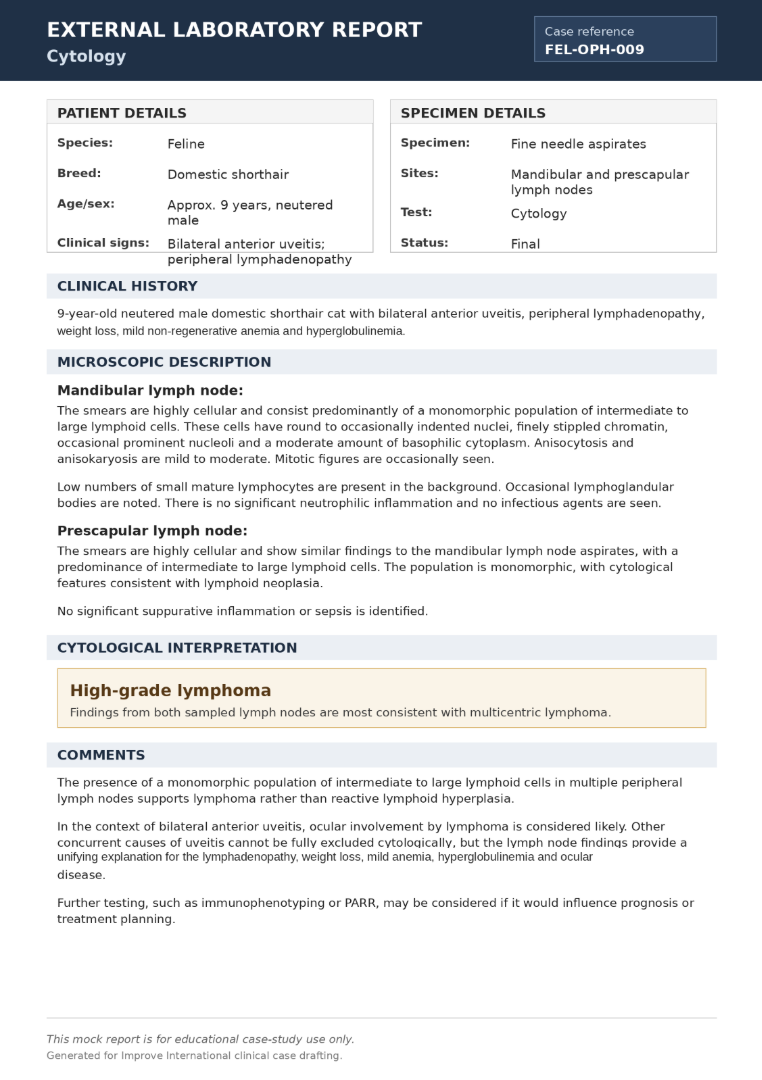
The cat now has a confirmed diagnosis of high-grade multicentric lymphoma, and the bilateral anterior uveitis is very likely to represent ocular involvement by lymphoma. The diagnosis explains the uveitis, but the immediate problem has not gone away: the right eye has developed secondary glaucoma.

  • Continue topical pressure-lowering treatment in the right eye and recheck IOP later the same day.
  • Continue topical NSAID therapy because active uveitis is still present.
  • Do not restart atropine in the right eye while IOP is raised; monitor the left eye closely because it remains uveitic.
  • Discuss systemic lymphoma treatment options, including oncology referral if the owner would consider chemotherapy.
  • Do not start systemic prednisolone automatically before the treatment discussion; if the owner chooses palliative care, it should be a deliberate decision.
  • Be realistic about comfort: if the right eye cannot be kept comfortable despite medical management, enucleation may become a welfare-focused option.

The key point is that cytology confirmed the diagnosis, but management still has two priorities: control the painful glaucomatous eye and plan treatment for systemic lymphoma.

Patient recheck exam

The next check of the patient in the evening shows the right eye has improved to 20-22 mmHg on topical dorzolamide/timolol, although mild corneal haze and anterior chamber flare persist. The left eye remains 10-12 mmHg. The cat is more comfortable and has eaten a small amount. You decide the cat can go overnight without a recheck.

See more See less


The following morning...

The following morning, the cat is even more comfortable. The IOP in the right eye is 18 mmHg, and the left eye remains 11 mmHg. It is time to phone the owner with an update on her cat and a management plan. You will also need to talk her through the recent results, and the next steps of investigation.

What further investigations will you offer now?

Further investigations are now aimed at staging the lymphoma, assessing fitness for treatment, and giving the owner a realistic discussion about prognosis, cost and options.

  • Thoracic imaging: offer three-view thoracic radiographs to look for mediastinal widening, thoracic lymphadenopathy or pulmonary involvement. Thoracic imaging is also useful before chemotherapy or if sedation/anesthesia might be needed later.
  • Abdominal ultrasound: assess abdominal lymph nodes, liver, spleen, kidneys and gastrointestinal tract. Ultrasound may identify additional sites of lymphoma and can help distinguish multicentric disease from concurrent alimentary involvement.
  • Further cytology or biopsy if needed: lymph node cytology is already sufficient to support high-grade lymphoma, but additional sampling can be considered if the distribution of disease is unclear or if it would change treatment planning.
  • Immunophenotyping and/or PARR: discuss whether further characterization would alter treatment choices or prognosis. Immunophenotyping may help distinguish B-cell from T-cell lymphoma; PARR can support clonality where diagnosis is uncertain.
  • Bone marrow assessment only if indicated: this is not a routine first step in general practice, but may be considered if there are unexplained cytopenias, circulating atypical cells or an oncologist recommends it.
  • Referral versus general practice management: oncology referral allows formal staging, protocol selection and prognosis discussion, but palliative care in general practice remains valid if referral is not possible or not desired.

The owner should understand that staging is not just more tests. It helps answer three practical questions: how widespread the lymphoma appears to be, whether the cat is well enough for treatment, and what treatment options are realistic.

In this case, the owner opts to undertake thoracic and abdominal imaging to stage the lymphoma.

See more See less

Test results

Thoracic radiographs

Three-view thoracic radiographs are obtained.

Finding

Result

Lung fields

No focal pulmonary mass or obvious metastatic pulmonary pattern

Pleural space

No pleural effusion

Cardiac silhouette

Within expected limits

Mediastinum

Mild cranial mediastinal widening

Thoracic lymph nodes

Sternal lymph node mildly enlarged; cranial mediastinal lymphadenopathy suspected

Other findings

No evidence of trauma or pneumonia

Radiographic interpretation: Mild thoracic lymphadenopathy is present. There is no pleural effusion or obvious pulmonary involvement.

Abdominal ultrasound

Abdominal ultrasound is performed the same day.

Structure

Finding

Liver

Mildly enlarged with subjectively mildly hypoechoic, uniform parenchyma

Spleen

Mildly enlarged with diffusely mottled echotexture

Abdominal lymph nodes

Moderate enlargement of jejunal and medial iliac lymph nodes; nodes are rounded and hypoechoic

Gastrointestinal tract

Mild, diffuse small intestinal wall thickening; layering largely preserved

Kidneys

Normal size and architecture; no renal mass identified

Bladder

Unremarkable

Peritoneal cavity

No abdominal effusion

Ultrasound interpretation: The findings are consistent with disseminated lymphoma, with abdominal lymphadenopathy and likely splenic and hepatic involvement. Mild intestinal wall thickening may represent lymphomatous involvement or concurrent inflammatory change.

Additional ultrasound-guided aspirates of the spleen, liver or abdominal lymph nodes could be considered, but the owner declines further sampling. The peripheral lymph node cytology has already confirmed high-grade lymphoma, and the purpose of the imaging was to guide the treatment and prognosis discussion rather than obtain another diagnosis.

Open results See less


Treatment planning

You relay the latest test results to the owner, and present her with some treatment options. 

What are the treatment options for this disease? What prognosis does each treatment option carry? 

Option

What it involves

Likely outcome/prognosis

Chemotherapy: full or less intensive protocols

Referral to oncology or chemotherapy in general practice if the practice is experienced with oncology protocols. Options may include COP, CHOP-type protocols or modified/single-agent approaches (Lomustine) depending on drug availability, comorbidities and owner's goals. Ocular treatment continues alongside systemic treatment.

Best chance of remission and meaningful survival time, but prognosis is still guarded because this is high-grade, disseminated disease with ocular involvement. Some cats have months to occasionally longer than a year; less intensive protocols may reduce burden but are generally less predictable.

Palliative prednisolone with ocular management

Systemic prednisolone with ongoing topical treatment for uveitis/glaucoma, analgesia and monitoring. Enucleation of the right eye can be discussed if the eye becomes blind and painful despite medical management, but this is a comfort procedure, not cancer treatment.

May temporarily improve appetite, comfort and lymph node size, but is not expected to produce durable remission. Survival is often weeks to a few months, depending on response, systemic progression and ocular comfort.

Euthanasia or comfort-focused endpoint

Appropriate if the owner does not want active treatment, if ocular pain cannot be controlled, if systemic signs progress, or if medication burden becomes unacceptable.

Prognosis without treatment is poor. The focus is avoiding prolonged discomfort, particularly from glaucoma or systemic decline.


 The owner listens to the options and opts for palliative treatment with prednisolone, ongoing medical management of the eyes, and euthanasia when deterioration occurs.  

See options See less

Write a go-home treatment plan for this cat, including doses and frequency of rechecks.  

Medication plan

For this 4.1 kg cat, a reasonable go-home plan would be:

Medication

Dose and route

Frequency

Notes

Prednisolone

10 mg PO

Once daily

Palliative treatment for lymphoma. Review after 5-7 days and reduce only if clinically appropriate. Do not combine with systemic NSAIDs. Use concurrent topical NSAID/steroid treatment only with a clear ophthalmic plan.

Flurbiprofen sodium 0.03% ophthalmic solution

1 drop into each eye

Every 8 hours initially

Continue while active anterior uveitis is present. Frequency can be reduced once flare, fibrin and comfort improve, but do not taper without rechecking the eyes and IOP.

Dorzolamide/timolol ophthalmic drops

1 drop into the right eye

Every 12 hours

Continue for the eye that developed secondary glaucoma. Recheck IOP before reducing or stopping. Avoid if significant bradycardia, asthma or cardiorespiratory concerns develop.

Buprenorphine

0.02 mg/kg oral transmucosal; for this cat, approximately 0.08 mg, which is about 0.25-0.3 mL of a 0.3 mg/mL preparation

Every 8-12 hours as needed for pain

Continue for the first 48-72 hours, then reassess. Use longer if ocular pain persists and the cat tolerates it well.

Atropine 1% ophthalmic ointment

No further atropine in the right eye. Consider atropine into the left eye only if still markedly miotic or painful

No more than once daily, and only as directed

Avoid routine ongoing use because both eyes are at risk of IOP changes. Stop if the left pupil is dilated, if the eye becomes more painful, or if IOP rises.

If the cat is difficult to medicate, prioritize systemic prednisolone, right-eye glaucoma medication, topical anti-inflammatory treatment, then analgesia as needed. If the medication burden itself is causing distress, that should form part of the quality-of-life discussion.

Recheck plan

  • 24 hours after discharge: check comfort, appetite, owner ability to medicate, and perform an ophthalmic recheck including IOP in both eyes.
  • 48-72 hours later: repeat ocular examination and IOP; review appetite, weight, hydration and lymph node size.
  • Weekly for the first 2-3 weeks if stable: monitor IOP, ocular comfort, uveitis, body weight, appetite, demeanor and lymph node size.
  • Then every 2-4 weeks if still comfortable, with earlier review for any ocular recurrence or systemic decline.

Signs that require urgent reassessment

  • the right eye becoming more closed, cloudy, red or painful, or a larger/fixed pupil developing
  • sudden change in vision, bumping into objects, pawing at the face or marked light sensitivity
  • reduced appetite for more than 24 hours, hiding, marked lethargy or withdrawal
  • difficulty giving medication, vomiting, diarrhea, dehydration, labored breathing, open-mouth breathing or collapse

Signs that euthanasia should be considered

  • persistent ocular pain or recurrent/uncontrolled glaucoma, especially if the eye is blind or likely to be blind
  • inability to keep the cat comfortable without stressful or frequent medication
  • anorexia or minimal intake despite palliative treatment, progressive weight loss, weakness or dehydration
  • respiratory difficulty, collapse, severe lethargy, hiding, loss of normal interaction or repeated bad days outnumbering good days

If the right eye becomes blind and painful but the cat is otherwise doing well, enucleation can be discussed as a comfort procedure. Given the disseminated lymphoma, this should be weighed against expected survival time, anesthetic burden, cost and the owner's goals.

The plan should be recorded clearly: this is palliative care, with active treatment for comfort, not curative treatment. The agreed endpoint is euthanasia when ocular pain, systemic decline or medication burden can no longer be managed in a way that preserves the cat’s quality of life.

See more See less


Key learning points

Once you have completed the case, consider what you've learned. This reflection is an important part of the CE cycle. Is there anything you'd do differently in future? Have you identified any knowledge gaps that need filling?

Key learning points to take away

Spoiler alert: do not open until you've worked through the whole case!

  • A bilateral red eye with discharge is not always conjunctivitis; miosis, photophobia, aqueous flare and low IOP should redirect the case toward anterior uveitis.
  • Bilateral anterior uveitis in a cat should trigger a systemic work-up, particularly when accompanied by weight loss, anemia, hyperglobulinemia or lymphadenopathy.
  • Atropine can complicate interpretation of pupil size. Repeat IOP is essential when a uveitic eye becomes more painful or cloudy.
  • Secondary glaucoma is an ocular emergency even when the underlying systemic diagnosis is still pending.
  • Lymphoma can present through the eyes, but treatment planning still needs to address both systemic disease and ocular comfort.
Open See less
IVE_GPCert(Ophthal)_OL_CourseThumb (3)
online

Ophthalmology

ISVPS General Practitioner Certificate (GPCert)

Postgraduate Certificate (PgC)

date Sept 2026 location Online